Date:
Copyright:
TAIJIRO TOMIKAWA, KAZUO SHIN-YA, HARUO SETO, NORIYUKI OKUSA, TAKAYUKI KAJIURA, YOICHI HAYAKAWA, Structure of Aspochalasin H, a New Member of the Aspochalasin Family, The Journal of Antibiotics, 2002, Volume 55, Issue 7, Pages 666-668, Released on J-STAGE January 27, 2009, Online ISSN 1881-1469, Print ISSN 0021-8820, https://doi.org/10.7164/antibiotics.55.666, https://www.jstage.jst.go.jp/article/antibiotics1968/55/7/55_7_666/_article/-char/en
Notes:
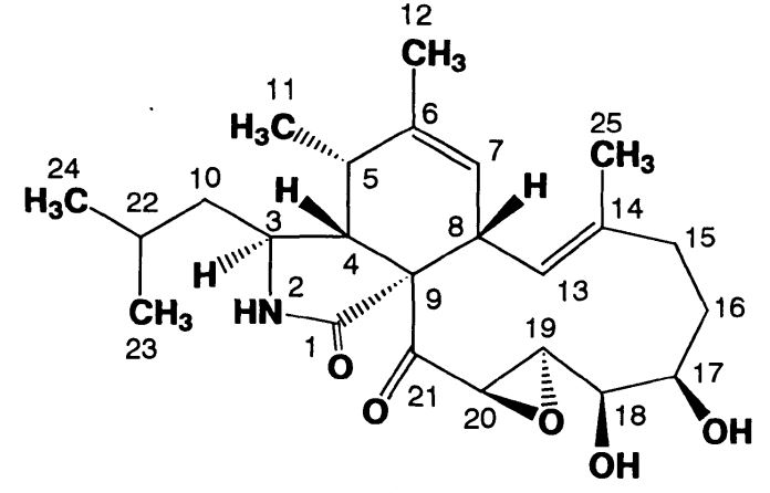
Structure of Aspochalasin H
Images library
-
Title
Legend
-
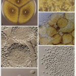
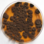
Pigmentation of Aspergillus versicolor colonies ranged from pale green to greenish-beige, pink-green, dark green and brown. Reverse is usually reddish. The growth rate is usually slow. Cultured on Sabouraud dextrose agar with chloramphenicol.
-
A Colonies on MEA after one week; B, C conidial heads with tip of conidiophire, x920; D conidial head, x 2330; E conidial heads x920
![aspvers[2] aspvers2](https://www.aspergillus.org.uk/wp-content/uploads/2017/10/aspvers2-150x150.jpg)
-
A Colonies on MEA + 20% sucrose after one week; B detail of colony showing columnar conidial heads x 44 ; C conidial heads x 920; D conidia x2330
-
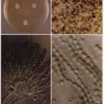
Cultures are grown on malt extract agar for 5-7 days at 30°C.
Light microscopy-1000x stained with lacto-phenol and cotton blue.
-
A Colonies on MEA +20% sucrose after one week; B ascomata x 40; C conidiophores x 920; D ascospores x2330; E ascoma x 230; F portion of ascoma with asci and ascospores, x 920.
