Date:
Copyright:
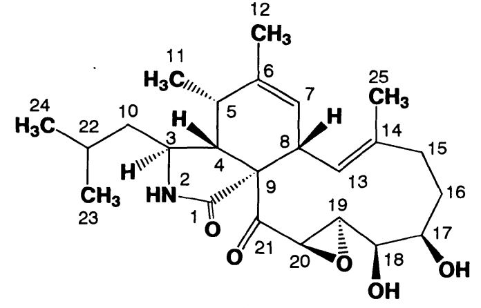
TAIJIRO TOMIKAWA, KAZUO SHIN-YA, HARUO SETO, NORIYUKI OKUSA, TAKAYUKI KAJIURA, YOICHI HAYAKAWA, Structure of Aspochalasin H, a New Member of the Aspochalasin Family, The Journal of Antibiotics, 2002, Volume 55, Issue 7, Pages 666-668, Released on J-STAGE January 27, 2009, Online ISSN 1881-1469, Print ISSN 0021-8820, https://doi.org/10.7164/antibiotics.55.666, https://www.jstage.jst.go.jp/article/antibiotics1968/55/7/55_7_666/_article/-char/en
Notes:
Structure of Aspochalasin H
Images library
-
Title
Legend
-
1 Axial computed tomography (CT) scans of the frontal sinus.
A: due to the long lasting pressure of mucus, the bone of the anterior wall of frontal sinus is thinned out and elevated anteriorly, forming a bulge. B: same situation as depicted in fig A: the posterior bony wall of frontal sinus is thinned out and extremely elevated posteriorly towards the frontal lobe of the brain. As depicted on the scan, a thin bony layer covering the dura could be recognized intraoperatively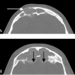
-
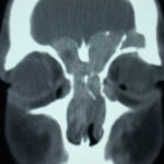
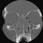
2 Same patient as 1 and 3, frontal CT
-
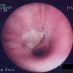
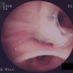
D. 6 months later, tenacious yellow secretions in L basal bronchial division
-
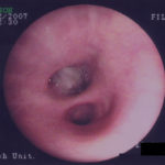
C. After suction the material was seen to extend distally – obstructing the right basal stem bronchus
-
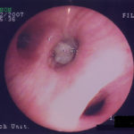
B. After suction the material was seen to extend distally – obstructing the right basal stem bronchus
-
A. Necrotic mass prolapsing in and out of the distal right intermediate bronchus obscuring both the basal stem and basal division
