Date:
Copyright:
FANG FANG, HIDEAKI UI, KAZURO SHIOMI, ROKURO MASUMA, YUUICHI YAMAGUCHI, CHENG GANG ZHANG, XIAN WU ZHANG, YOSHITAKE TANAKA, SATOSHI OMURA, Two New Components of the Aspochalasins Produced by Aspergillm sp., The Journal of Antibiotics, 1997, Volume 50, Issue 11, Pages 919-925, Released on J-STAGE November 25, 2006, Online ISSN 1881-1469, Print ISSN 0021-8820, https://doi.org/10.7164/antibiotics.50.919, https://www.jstage.jst.go.jp/article/antibiotics1968/50/11/50_11_919/_article/-char/en
Notes:
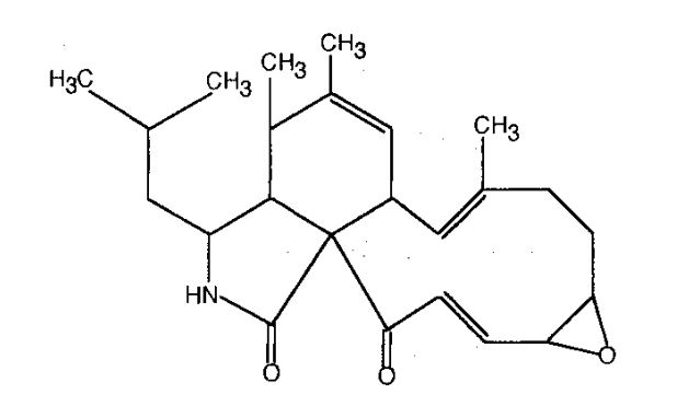
Structure of aspochalasin G
Images library
-
Title
Legend
-
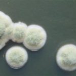
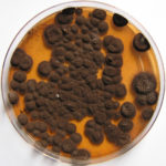
Pigmentation of Aspergillus versicolor colonies ranged from pale green to greenish-beige, pink-green, dark green and brown. Reverse is usually reddish. The growth rate is usually slow. Cultured on Sabouraud dextrose agar with chloramphenicol.
-
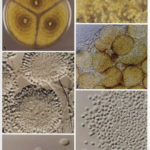
A Colonies on MEA after one week; B, C conidial heads with tip of conidiophire, x920; D conidial head, x 2330; E conidial heads x920
![aspvers[2] aspvers2](https://www.aspergillus.org.uk/wp-content/uploads/2017/10/aspvers2-150x150.jpg)
-
A Colonies on MEA + 20% sucrose after one week; B detail of colony showing columnar conidial heads x 44 ; C conidial heads x 920; D conidia x2330
-
Cultures are grown on malt extract agar for 5-7 days at 30°C.
Light microscopy-1000x stained with lacto-phenol and cotton blue.
-
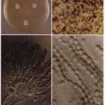
A Colonies on MEA +20% sucrose after one week; B ascomata x 40; C conidiophores x 920; D ascospores x2330; E ascoma x 230; F portion of ascoma with asci and ascospores, x 920.
