Date: 26 November 2013
Further details
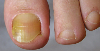
Image 5. Oral itraconazole pulse therapy was given to the patient (200 mg twice daily for 1 week, with 3 weeks off between successive pulses, for four pulses) and treatment was successful.
Copyright:
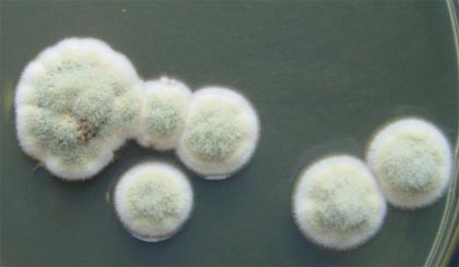
Image 1. Copyright Fungal Research Trust.
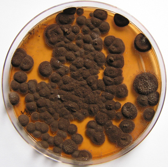
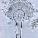
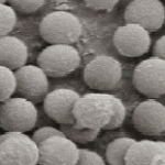
Image 2. Copyright B.Flannigan, R Samson & JD Miller (From Microorganisms in home and indoor work environments, Published by Taylor and Francis)
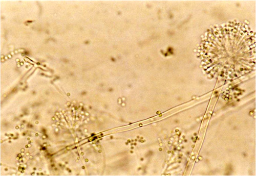
Images 3-5. With thanks to S Veraldi, A Chiaratti and H Harak Institute of Dermatological Sciences, University of Milan. Italy . These images remain the copyright of ‘Mycoses’ where the full article may be viewed. (Veraldi et al, published online Mycoses, 5th May 2009 http://www3.interscience.wiley.com/cgi-bin/fulltext/122374087/HTMLSTART).
Notes: n/a
Images library
-
Title
Legend
-
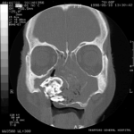
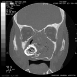
Patient MB X rays and CT scans. Chronic calcified maxillary sinusitis, patient had a palate defect.A. fumigatus cultured.
Images A&B Plain X rays antero-posterior and lateral, pre-operatively of Pt MB aged 76 who presented with unilateral nasal stuffiness and difficulty getting dentures fitted. She had hda these symptoms for many years. A large irregular calcified mass can be seen replacing the right maxillary sinus.
Images C D & E Coronal CT scan images of Pt MB showing a completely obstructed nasal cavity bilaterally and loss of internal nasal architecture. On the right side is large lamellar calcified lesion embedded in the extensive inflammatory material. Loss of bony margins is seen in numerous locations. This material was all removed surgically and showed mostly necrotic debris with Charcot-Leyden crystals and a few eosinophils and degenerate fungal hyphae. Aspergillus fumigatus was cultured from the material, especially infero-laterally on the right.
Image F Photograph through the mouth post-operatively showing the palate and a large defect in its right side. Through the defect can be seen the interior of the right maxillary sinus and nasal cavity with the inferior turbinate just visible.
 ,
,  ,
,  ,
,  ,
,  ,
, 

