Date: 8 January 2014
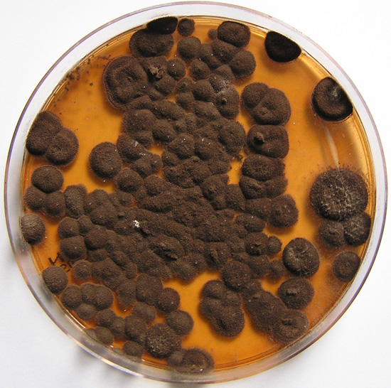
Pigmentation of Aspergillus versicolor colonies ranged from pale green to greenish-beige, pink-green, dark green and brown. Reverse is usually reddish. The growth rate is usually slow. Cultured on Sabouraud dextrose agar with chloramphenicol.
Copyright:
With thanks to S Veraldi, A Chiaratti and H Harak Institute of Dermatological Sciences, University of Milan. Italy . These images remain the copyright of ‘Mycoses’ where the full article may be viewed. (Veraldi et al, published online Mycoses, 5th May 2009).
Notes: n/a
Images library
-
Title
Legend
-
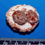
Gross pathology demonstrating the great pleural thickness and two cavities (upper lobe and superior segment of lower lobe) with fragments of fungal mass.

-
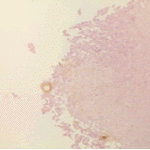
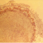
Histopathological appearance of a fungus ball. Note a conidial head resulting from fungal exposure to the air.
-
Histopathological appearance of a fungus ball caused by Scedosporium apiospermum. The presence of anneloconidia differentiates it from Aspergillus.

-
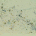
Chronic necrotising aspergillosis. Hyaline hyphal and calcium oxalate crystals obtained by needle aspirate biopsy from a diabetic patient with chronic necrotizing aspergillosis caused by Aspergillus niger (Papanicolaou, x 100).
-
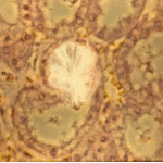
Aspergillus niger fungus ball and acute oxalosis. Higher magnification of adjacent replicate section.
-
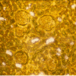
Oxalate crystals within renal tubuli (H&E, phase contrast, x 100). This patient developed acute oxalosis.
-
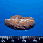
Lung surface. Fungus ball, severe parenchymal fibrosis and pleural thickening.

-
The periphery of the fungus ball is deeply eosinophilic because of the deposition of Splendore-Hoeppli material.

 ,
, 
 ,
,  ,
, 