Date: 8 January 2014
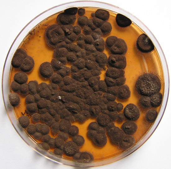
Pigmentation of Aspergillus versicolor colonies ranged from pale green to greenish-beige, pink-green, dark green and brown. Reverse is usually reddish. The growth rate is usually slow. Cultured on Sabouraud dextrose agar with chloramphenicol.
Copyright:
With thanks to S Veraldi, A Chiaratti and H Harak Institute of Dermatological Sciences, University of Milan. Italy . These images remain the copyright of ‘Mycoses’ where the full article may be viewed. (Veraldi et al, published online Mycoses, 5th May 2009).
Notes: n/a
Images library
-
Title
Legend
-
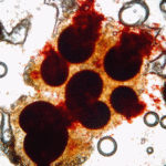
Emericella nidulans (Eidam), Anamorph: Aspergillus nidulans (Eidam) – Hulle cells

-
Emericella nidulans (Eidam), Anamorph: Aspergillus nidulans (Eidam)

-
Emericella nidulans (Eidam), Anamorph: Aspergillus nidulans (Eidam)

-
Emericella nidulans (Eidam), Anamorph: Aspergillus nidulans (Eidam)

-
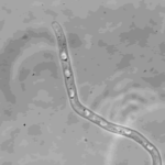
Spitzenkorper; phase contrast micrograph of A.nidulans after one day grown at 25°C in minimal media containing 17% gelatin showing spitzenkorper and fungal cell morphology
-
Interference contrast micrograph of A.nidulans after one day at 25°C in minimal media containing 17% gelatin

-
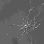
Hyphal growth. Micro-colony of A.nidulans grown overnight at 25°C in minimal media containing 17% gelatin
-
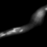
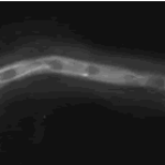
Mitochondria organisation: GFP fluorescence micrographs showing mitochondrial organisation in an A.nidulans strain with GFP mitochondria, grown at 25°C in minimal media.
