Date: 8 January 2014
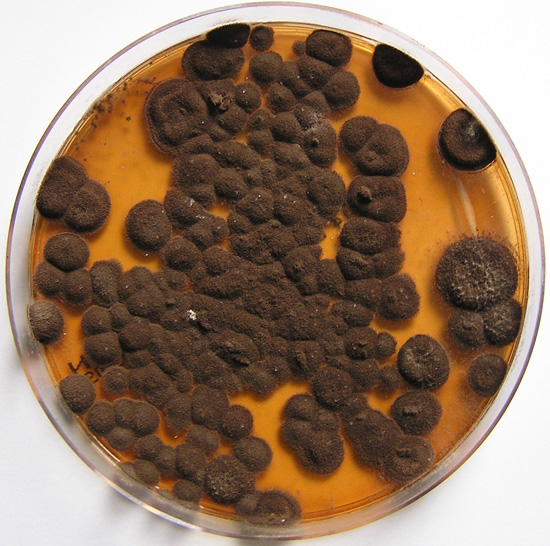
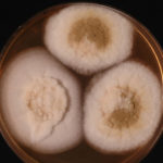
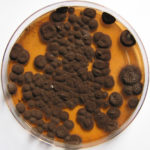
Pigmentation of Aspergillus versicolor colonies ranged from pale green to greenish-beige, pink-green, dark green and brown. Reverse is usually reddish. The growth rate is usually slow. Cultured on Sabouraud dextrose agar with chloramphenicol.
Copyright:
With thanks to S Veraldi, A Chiaratti and H Harak Institute of Dermatological Sciences, University of Milan. Italy . These images remain the copyright of ‘Mycoses’ where the full article may be viewed. (Veraldi et al, published online Mycoses, 5th May 2009).
Notes: n/a
Images library
-
Title
Legend
-
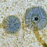
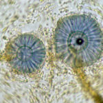
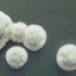
Scanning electron micrograph of Aspergillus ochraceopetaliformis conidial heads
-
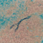
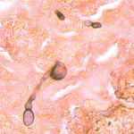
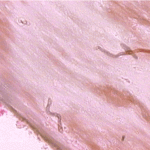
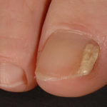
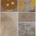
Image D & E. A case of onychomycosis associated with Aspergillus ochraceopetaliformis as described in Nail infection by Aspergillus ochraceopetaliformis. Med Mycol. 2009 Mar 9:1-5, 2009, Brasch J, Varga J, Jensen JM, Egberts F & Tintelnot K
 ,
,  ,
,  ,
,  ,
, 
-
Further details
Image 5. Oral itraconazole pulse therapy was given to the patient (200 mg twice daily for 1 week, with 3 weeks off between successive pulses, for four pulses) and treatment was successful.
 ,
,  ,
,  ,
,  ,
, 
-
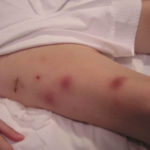
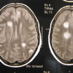
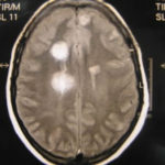
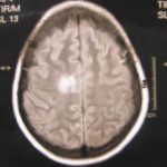
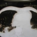
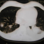
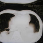
This patient was 28 yr old with adult lymphocytic leukaemia. She received induction chemotherapy and this infection developed 2 days after recovering from neutropenia.
 ,
,  ,
,  ,
,  ,
,  ,
,  ,
,  ,
,  ,
,  ,
, 
-
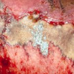
Close-up image of the lesion on the left thigh showing a mat of hyphae over the wound.
-
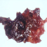
Eosinophilic mucin with A. flavus in the nasal cavity. Irregular crust of 2.5 cm from a patient diagnosed as allergic fungal sinusitis. Patient with allergic fungal sinusitis
-
GMS stain of eosinophilic mucin reveals a darkly stained dichotomously branched A. flavus hyphae within cellular background. Patient with allergic fungal sinusitis
