Date: 26 November 2013
Conidial heads from culture on CYA 25°C medium (mag x100)
Copyright:
These images were generously provided by Mirca Zotti, University of Genoa who retains the Copyright
Notes: n/a
Images library
-
Title
Legend
-
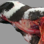
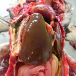
Aspergillosis in penguins. Lesions found in captive Magellanic penguins (Spheniscus magellanicus) with aspergillosis as determined by histology. Adherence areas of air sac to the celomic wall and white-yellowish nodule in the liver.

-
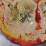
Aspergillosis in penguins. Lesions found in fatal cases of captive Magellanic penguins (Spheniscus magellanicus) with aspergillosis. Air sacs thickened with abundant plaque-like caseous and necrotic debris covering the wall with greyish-green fungal colonies on the internal surface.
-
Aspergillosis in penguins. Lesions found in fatal cases of captive Magellanic penguins (Spheniscus magellanicus) with aspergillosis. Air sacs thickened with abundant plaque-like caseous and necrotic debris covering the wall with greyish-green fungal colonies on the internal surface.
-
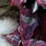
Aspergillosis in penguins. Lesions found in fatal cases of captive Magellanic penguins (Spheniscus magellanicus) with aspergillosis. Air sacs thickened with abundant plaque-like caseous and necrotic debris covering the wall with greyish-green fungal colonies on the internal surface.
-
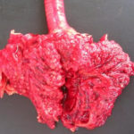
Aspergillosis in penguins. Lesions found in captive Magellanic penguins (Spheniscus magellanicus) with aspergillosis.Lung parenchyma with congestion, hemorrhagic and necrotic areas and with multiple white-yellowish granulomatous nodules, ranging from 0.1-1.0 cm in diameter

-
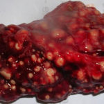
Aspergillosis in penguins. Lesions found in captive Magellanic penguins (Spheniscus magellanicus) with aspergillosis. Lung parenchyma with congestion, hemorrhagic and necrotic areas and with multiple white-yellowish granulomatous nodules, ranging from 0.1-1.0 cm in diameter
-
Aspergillosis in penguins. Lesions found in captive Magellanic penguins (Spheniscus magellanicus) with aspergillosis.
