Date: 26 November 2013
renal transplant patient
Copyright:
Kindly provided by Iván Solano Leiva, Infectious Diseases MD, Instituto Salvadoreno del Seguro Social, El Salvador.
Notes:
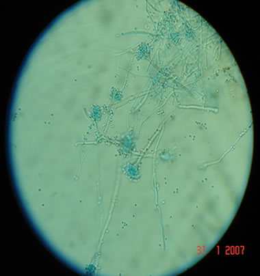
A 54 yr old male patient who underwent a renal transplant one year earlier. The patient noticed a lesion on left plantar region, it was not painful but was slowly enlarging (over 9 months). In the latter 3 months the lesion became slightly purulent. Multiple cycles of antibiotics gave no improvemnt. Culture of the discharge produced Aspergillus niger. The final diagnosis of this patient was Aspergillus osteomyelitis. The patient was treated with voriconazole orally 200 mg twice daily.
Images library
-
Title
Legend
-
Necrotic lung tissue in culture
Af=Colony of Aspergillus fumigatus
B=bacterial colonies
L=lung tissue
-
A photograph of part of the upper lung lobe of an immunosuppressed patient. The lung tissue shows extenive areas of necrosis due to invasive colonisation.
-
A photograph of a cavity in the upper lobe of the lung of a patient with ankylosing spondylitis. Such cavitation,which may be confused with prior tuberculosis, can follow fibrosis.
-
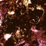
Plugs stained with Methenamine/silverActively growing mycelia of the fungus are a deep brown/black. Counterstaining shows the dense mucus of the plugs as predominantly orange.
-
Sputum from an asthmatic patient showing plugs(casts). The development of plugs coincided with an increased prevalence and severity of episodes of asthma

-
microscopic characters Conidiophore stipes(C)1300-2800um long:Vesicles(V)40-70um wide,clavate:Phialides(Ph) uniseriate:Conidia(Con)3.5-4.0um long,smooth walled.

-
microscopic characters Conidiophore stipes(C)225-350um arising from hyphae(Hy):Vesicles(Ves)15-25um wide:Phialides(Ph)uniseriate:Conidia(Con)2.4-3.0um spherical to ovoid,roughened.


