Date: 26 November 2013
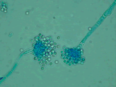
Light microscopy at 1000x stained with lacto-phenol cotton blue.
Copyright:
Images were kindly provided by Niall Hamilton Copyright Fungal Research Trust
Notes:
Distinctive features
Colony characteristics. Colonies (CzA) growing rather slowly, white.Microscopy. Cinidiophore stipes smooth walled, hyaline, up to 1000 micrometre long. Conidial heads small, radiate to loosely columnar, white, becoming dull ivory with age. Vesicles hemispherical, 8-15 micrometre diam. Conidiogenous cells biseriate. Metulae covering the upper one- to two-thirds of the vesicle. Conidia spherical, hyaline, 2-2.5 micrometre daim, smooth walled.
Images library
-
Title
Legend
-
Single fungal ball, moving. Radiographic appearance of a fungus ball, showing movement as the patient’s position changes.
-
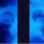
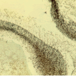
Oxalate crystals in the cavity wall surrounding an Aspergillus niger fungus ball (H&E, dark field, x 25).
-
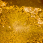
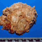
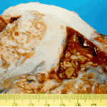
Aspergilloma patient. Gross pathology appearance of a fungus ball.
-
Conidiophores of Aspergillus fumigatus in the mass of the fungal ball surrounded by mycelia (H&E, x 400).
-
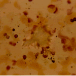
Aspergillus niger fungal ball. Calcium oxalate crystals in Aspergillus niger fungal ball. Also shown are darkly pigmented, rough-walled conidia associated with Aspergillus niger infection.
-
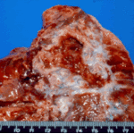
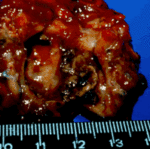
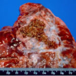
Aspergillus niger fungus ball within an old tuberculous cavern. This patient had diabetes, a disease commonly associated with A. niger infection.
-
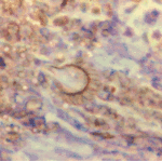
Conidial head and brown conidia in a section of a fungus ball caused by Aspergillus niger (H&E, x 400).


 ,
, 
 ,
, 