Date: 26 November 2013
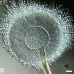
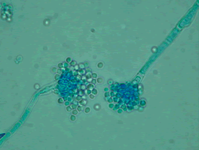
Light microscopy at 1000x stained with lacto-phenol cotton blue.
Copyright:
Images were kindly provided by Niall Hamilton Copyright Fungal Research Trust
Notes:
Distinctive features
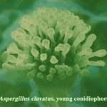
Colony characteristics. Colonies (CzA) growing rather slowly, white.Microscopy. Cinidiophore stipes smooth walled, hyaline, up to 1000 micrometre long. Conidial heads small, radiate to loosely columnar, white, becoming dull ivory with age. Vesicles hemispherical, 8-15 micrometre diam. Conidiogenous cells biseriate. Metulae covering the upper one- to two-thirds of the vesicle. Conidia spherical, hyaline, 2-2.5 micrometre daim, smooth walled.
Images library
-
Title
Legend
-
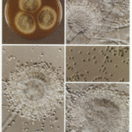
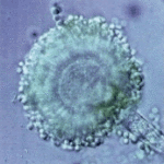
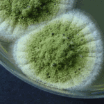
Aspergillus flavus Link. A colonies after 1 weekB,C conidial heads x 920D Conidia x920 E Conidial head x920
-
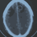
Patient diagnosed with Stage C Chronic Lymphocytic Leukaemia, treated in the MRC CLL4 study, with prednisolone for 4 weeks followed by oral chlorambucil for 7 days. Patient developed severe pneumonia due to pseudomonas and staphylococcus. Following treatment with broad spectrum antibiotics and 1 week of Abelcet, patient was readmitted with headache, disorientation and fever. CT brain scans showed 3 ring enhancing lesions, aspirated material showed neutrophils but grew aspergillus. Patient now improved on Abelcet (4 mg/kg) and oral itrconazole suspension 200mg b.d.
Thanks to Richard Chasty, Consultant Haematologist, North Staffordsire Hospital, UK.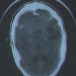 ,
, 

![Asp[1]niger Asp[1]niger](https://www.aspergillus.org.uk/wp-content/uploads/2013/11/Asp1niger-150x150.gif)


