Date: 26 November 2013
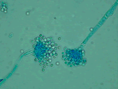
Light microscopy at 1000x stained with lacto-phenol cotton blue.
Copyright:
Images were kindly provided by Niall Hamilton Copyright Fungal Research Trust
Notes:
Distinctive features
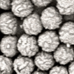
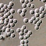
Colony characteristics. Colonies (CzA) growing rather slowly, white.Microscopy. Cinidiophore stipes smooth walled, hyaline, up to 1000 micrometre long. Conidial heads small, radiate to loosely columnar, white, becoming dull ivory with age. Vesicles hemispherical, 8-15 micrometre diam. Conidiogenous cells biseriate. Metulae covering the upper one- to two-thirds of the vesicle. Conidia spherical, hyaline, 2-2.5 micrometre daim, smooth walled.
Images library
-
Title
Legend
-
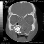
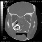
Patient MB X rays and CT scans. Chronic calcified maxillary sinusitis, patient had a palate defect.A. fumigatus cultured.
Images A&B Plain X rays antero-posterior and lateral, pre-operatively of Pt MB aged 76 who presented with unilateral nasal stuffiness and difficulty getting dentures fitted. She had hda these symptoms for many years. A large irregular calcified mass can be seen replacing the right maxillary sinus.
Images C D & E Coronal CT scan images of Pt MB showing a completely obstructed nasal cavity bilaterally and loss of internal nasal architecture. On the right side is large lamellar calcified lesion embedded in the extensive inflammatory material. Loss of bony margins is seen in numerous locations. This material was all removed surgically and showed mostly necrotic debris with Charcot-Leyden crystals and a few eosinophils and degenerate fungal hyphae. Aspergillus fumigatus was cultured from the material, especially infero-laterally on the right.
Image F Photograph through the mouth post-operatively showing the palate and a large defect in its right side. Through the defect can be seen the interior of the right maxillary sinus and nasal cavity with the inferior turbinate just visible.
 ,
,  ,
,  ,
,  ,
,  ,
, 
-
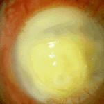
Aspergillus keratitis. Severe aspergillus infection with large area of corneal ulceration and deep stromal involvement

-
Sequence of images showing ocular surface change which unusually predisposed to severe fusarium keratitis in an elderly woman. Successful treatment involved full thickness corneal transplantation shown 2 weeks and then 2 years after surgery.

-
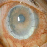
Sequence of images showing ocular surface change which unusually predisposed to severe fusarium keratitis in an elderly woman. Successful treatment involved full thickness corneal transplantation shown 2 weeks and then 2 years after surgery.
-
Sequence of images showing ocular surface change which unusually predisposed to severe fusarium keratitis in an elderly woman. Successful treatment involved full thickness corneal transplantation shown 2 weeks and then 2 years after surgery.
-
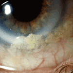
Sequence of images showing ocular surface change which unusually predisposed to severe fusarium keratitis in an elderly woman. Successful treatment involved full thickness corneal transplantation shown 2 weeks and then 2 years after surgery
-
Aspergillus keratitis. Shrunken eye as a consequence of this infection


