Date: 26 November 2013
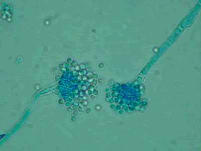
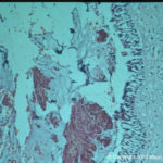
Light microscopy at 1000x stained with lacto-phenol cotton blue.
Copyright:
Images were kindly provided by Niall Hamilton Copyright Fungal Research Trust
Notes:
Distinctive features
Colony characteristics. Colonies (CzA) growing rather slowly, white.Microscopy. Cinidiophore stipes smooth walled, hyaline, up to 1000 micrometre long. Conidial heads small, radiate to loosely columnar, white, becoming dull ivory with age. Vesicles hemispherical, 8-15 micrometre diam. Conidiogenous cells biseriate. Metulae covering the upper one- to two-thirds of the vesicle. Conidia spherical, hyaline, 2-2.5 micrometre daim, smooth walled.
Images library
-
Title
Legend
-
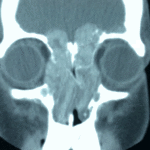
4 Total obstruction of the sinuses due to inflamed mucosa. (Patient 04)
-
1 Axial computed tomography (CT) scans of the frontal sinus.
A: due to the long lasting pressure of mucus, the bone of the anterior wall of frontal sinus is thinned out and elevated anteriorly, forming a bulge. B: same situation as depicted in fig A: the posterior bony wall of frontal sinus is thinned out and extremely elevated posteriorly towards the frontal lobe of the brain. As depicted on the scan, a thin bony layer covering the dura could be recognized intraoperatively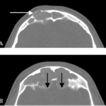
-
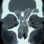
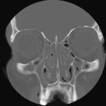
2 Same patient as 1 and 3, frontal CT
-
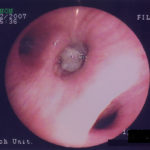
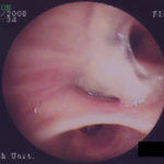
D. 6 months later, tenacious yellow secretions in L basal bronchial division
-
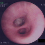
C. After suction the material was seen to extend distally – obstructing the right basal stem bronchus
-
B. After suction the material was seen to extend distally – obstructing the right basal stem bronchus
