Date: 26 November 2013
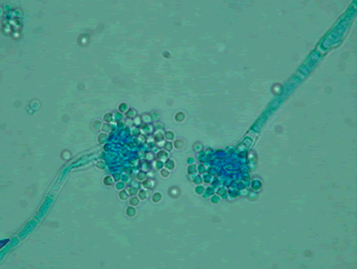
Light microscopy at 1000x stained with lacto-phenol cotton blue.
Copyright:
Images were kindly provided by Niall Hamilton Copyright Fungal Research Trust
Notes:
Distinctive features
Colony characteristics. Colonies (CzA) growing rather slowly, white.Microscopy. Cinidiophore stipes smooth walled, hyaline, up to 1000 micrometre long. Conidial heads small, radiate to loosely columnar, white, becoming dull ivory with age. Vesicles hemispherical, 8-15 micrometre diam. Conidiogenous cells biseriate. Metulae covering the upper one- to two-thirds of the vesicle. Conidia spherical, hyaline, 2-2.5 micrometre daim, smooth walled.
Images library
-
Title
Legend
-
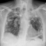
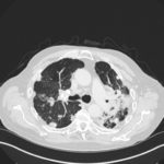
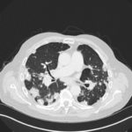
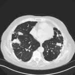
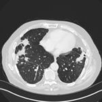
Mr RM is 80 and an ex-coal miner.He developed pneumoconiosis from exposure to coal dust. He also developed rheumatoid arthritis and the combination of this disease and pneumoconiosis is called Caplan’s syndrome.
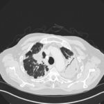
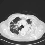
His chest Xray in early 2015 shows extensive bilateral pulmonary shadowing with solid looking nodules superimposed on abnormal lung fields, contraction of his left lung with an elevated diaphragm and a large left upper lobe aspergilloma, displaying a classic air crescent. His CT scan from mid 2014 demonstrates a large aspergilloma in a cavity on the left, with marked pleural thickening around it, which is partially ‘calcified’ towards its base. Inferiorly on other images,remarkable pleural thickening and fibrotic irregular and spiculated nodules are seen, most partially calcified.
 ,
,  ,
,  ,
,  ,
,  ,
,  ,
, 