Date: 7 May 2013
Copyright:
Copyright B.Flannigan, R Samson & JD Miller (From Microorganisms in home and indoor work environments, Published by Taylor and Francis)
Notes:
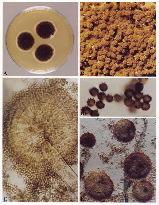
Colonies on CYA 60 mm or more diam, usually covering the whole Petri dish, plane, velutinous, of low, usually subsurface, white mycelium, surmounted by a layer of closely packed, black conidial heads, ca 2-3 mm high; reverse usually pale, sometimes pale to bright yellow. Colonies on MEA varying from 30-60 mm diam, usually smaller than those on CYA and often quite sparse by comparison, otherwise similar. Colonies on G25N 18-30 mm diam, plane, velutinous, with white or pale yellow mycelium visible at the margins, otherwise similar to those on CYA; reverse pale or occasionally with areas of deep brown. No growth at 5°C. At 37°C, colonies 60 mm or more diam, covering the available space, sometimes sulcate, otherwise similar to those on CYA at 25°C.
Conidiophores borne from surface hyphae, 1.0-3.0 mm long, with heavy, hyaline, smooth walls; vesicles spherical, usually 50-75 µm diam, bearing closely packed metulae and phialides over the whole surface; metulae 10-15 µm long, or sometimes more; phialides 7-10 µm long; conidia spherical, 4-5 µm diam, brown, with walls conspicuously roughened or sometimes striate, borne in large, radiate heads.
Distinctive features
One of the best known of all fungal species, Aspergillus niger is distinguished by its spherical black conidia, derived from colonies which show little or no other colouring.
Images library
-
Title
Legend
-
Image A
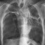
CT Scan 30/3/99
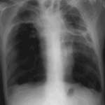
Showing extreme pleural thickening and 2 small cavities at apex of left lung.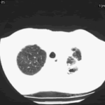
-
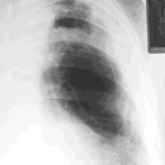
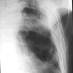
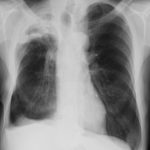
A 43 year old with smoking related emphysema was admitted to hospital with two separate episodes of haemoptysis. He had been in good health up to 1989, when he was diagnosed as having bilateral pulmonary tuberculosis. At that time a CT scan revealed a cavity in the left upper lobe (20.8cm2) with adjacent confluent infiltrates and pleural thickening. On bronchoscopic examination no abnormalities were noted and endobronchial biopsies did not reveal hyphae.
Over the next 4 years his condition deteriorated and a CT scan showed the left upper lobe cavity had increased to 40cm2. Itraconazole 400mg daily was prescribed. There was some clinical improvement on itraconazole but patient eventually deteriorated with breathlessness and with significant weight loss.
 ,
, 