Date: 26 November 2013
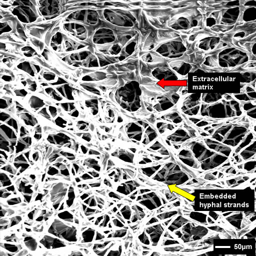
Biofilms on bronchial epithelial cells in vitro.
Copyright:
Images kindly donated by Frank-Michael C. Muller, Pediatric Pulmonology, Cystic Fibrosis Centre and Infectious Diseases, Department of Pediatrics III, University of Heidelberg, Im Neuenheimer Feld 430, D-69120 Heidelberg, Germany. © Fungal Research Trust
Notes:
An SEM of A. fumigatus of biofilm surface on bronchial epithelial cells at 48 h showing parallel-packed hyphae and ECM embedded in the hyphal network.
Images library
-
Title
Legend
-
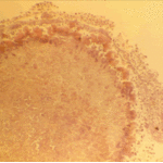
The periphery of the fungus ball is deeply eosinophilic because of the deposition of Splendore-Hoeppli material.
-
Single fungal ball, moving. Radiographic appearance of a fungus ball, showing movement as the patient’s position changes.
-
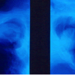
Oxalate crystals in the cavity wall surrounding an Aspergillus niger fungus ball (H&E, dark field, x 25).
-
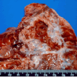
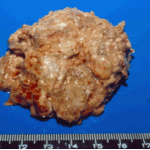
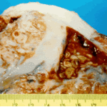
Aspergilloma patient. Gross pathology appearance of a fungus ball.
-
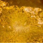
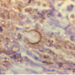
Conidiophores of Aspergillus fumigatus in the mass of the fungal ball surrounded by mycelia (H&E, x 400).
-
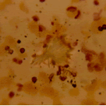
Aspergillus niger fungal ball. Calcium oxalate crystals in Aspergillus niger fungal ball. Also shown are darkly pigmented, rough-walled conidia associated with Aspergillus niger infection.
-
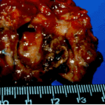
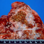
Aspergillus niger fungus ball within an old tuberculous cavern. This patient had diabetes, a disease commonly associated with A. niger infection.

 ,
, 
 ,
, 