Date: 26 November 2013
Biofilms on bronchial epithelial cells in vitro.
Copyright:
Images kindly donated by Frank-Michael C. Müller, Pediatric Pulmonology, Cystic Fibrosis Centre and Infectious Diseases, Department of Pediatrics III, University of Heidelberg, Im Neuenheimer Feld 430, D-69120 Heidelberg, Germany.
Notes:
Confocal scanning laser microscopy (CLSM), using CAAF(green) Fun1(red) stained biofilm. The red color of the FUN 1 cell stain was localized in dense aggregates in the cytoplasm of metabolically active cells. Thus, areas of red fluorescence represented metabolically active cells, and green fluorescence indicated cell wall-like polysaccharides, while yellow areas represented dual staining.
Images library
-
Title
Legend
-
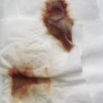
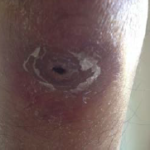
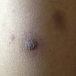
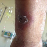
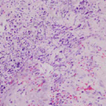
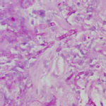
Images and abstract taken from Mert D et.al., Hematol Rep. 2017 Jun 1;9(2):6997. doi: 10.4081/hr.2017.6997. Invasive Aspergillosis with Disseminated Skin Involvement in a Patient with Acute Myeloid Leukemia: A Rare Case.
Invasive pulmonary aspergillosis is most commonly seen in immunocompromised patients. Besides, skin lesions may also develop due to invasive aspergillosis in those patients. A 49-year-old male patient was diagnosed with acute myeloid leukemia.
The patient developed bullous and zosteriform lesions on the skin after the 21st day of hospitalization. The skin biopsy showed hyphae. Disseminated skin aspergillosis was diagnosed to the patient.
Voricanazole treatment was initiated. The patient was discharged once the lesions started to disappear.
 ,
,  ,
,  ,
,  ,
, 
-
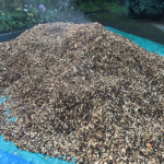
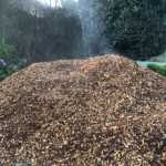
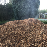
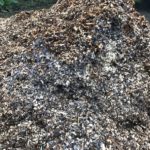
A pile of woodchip stored for use in a garden usually as a weed suppressing mulch. The heat building up in the pile is illustrated by the plumes of steam eminating from the top of the pile.
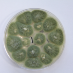
Aspergillus fumigatus is particularly well adapted to grow in the heat (up to 60C) found in such piles of rotting organic material and this characteristic, an adaption for its life in its natural environment also enables it to survive and grow in warm mammalian bodies at 37C. Most fungi cannot grow or survive at those temperatures
 ,
,  ,
,  ,
, 
-
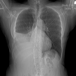
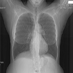
MK is 59 years old and presented with right sided pleuritic chest pain and coughing over 1 week. A chest Xray and then CT scan revealed complete collapse of her right lower lobe and middle lobes. Mucous retention is seen just proximal to the abrupt cutoff. There was mild bronchiectasis.
 ,
, 


 ,
,  ,
, 