Date: 26 November 2013
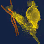
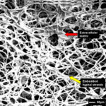
Biofilms on bronchial epithelial cells in vitro.
Copyright:
Images kindly donated by Frank-Michael C. Müller, Pediatric Pulmonology, Cystic Fibrosis Centre and Infectious Diseases, Department of Pediatrics III, University of Heidelberg, Im Neuenheimer Feld 430, D-69120 Heidelberg, Germany.
Notes:
Confocal scanning laser microscopy (CLSM), using CAAF(green) Fun1(red) stained biofilm. The red color of the FUN 1 cell stain was localized in dense aggregates in the cytoplasm of metabolically active cells. Thus, areas of red fluorescence represented metabolically active cells, and green fluorescence indicated cell wall-like polysaccharides, while yellow areas represented dual staining.
Images library
-
Title
Legend
-
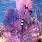
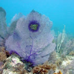
Aspergillosis in the coral sea fan Gorgonia ventalina. Sea fan coral Gorgonia ventalina, Florida Keys, USA. Depth ~5 metres, showing a lesion surrounded by a band of purple tissue. Central areas of the lesion are devoid of coral tissue revealing the underlying axial skeleton. The purple areas are devoid of coral polyps and result from the increased production of purple sclerites (small, non-fused, carbonate skeletal elements). The purpled area also indicates the location of high fungal hyphal density and el
-
Aspergillosis in the coral sea fan Gorgonia ventalina. In the Caribbean ond offshore USA the sea fan coral species Gorgonia ventalina is undergoing an epizootic due to Aspergillus sydowii infection. This species of Aspergillus is also known to be associated with food contamination and for opportunistic infection in humans.Taken in San Salvador, Bahamas. Bar represents 5cm. This sea fan was heavily colonized by algae.A -tumours and galls B -lesion C -purplingGalls are composed of axial skeleton and scl
