Date: 26 November 2013
Biofilms on bronchial epithelial cells in vitro.
Copyright:
Images kindly donated by Frank-Michael C. Müller, Pediatric Pulmonology, Cystic Fibrosis Centre and Infectious Diseases, Department of Pediatrics III, University of Heidelberg, Im Neuenheimer Feld 430, D-69120 Heidelberg, Germany.
Notes:
Confocal scanning laser microscopy (CLSM), using CAAF(green) Fun1(red) stained biofilm. The red color of the FUN 1 cell stain was localized in dense aggregates in the cytoplasm of metabolically active cells. Thus, areas of red fluorescence represented metabolically active cells, and green fluorescence indicated cell wall-like polysaccharides, while yellow areas represented dual staining.
Images library
-
Title
Legend
-
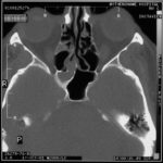
Allergic Aspergillus Sinusitis -Patient AM. D – Marked involvement of ehmoidal air cells on the right , together with the inferior aspect of the sphenoid sinus. The left side is almost clear of disease.
-
Further details
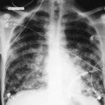
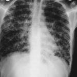
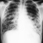
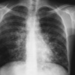
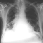
Image 1. The chest x-ray shows extensive bilateral nodular disease, most consistent with a fungal infection, or possibly tuberculosis. He was treated with a bucket face mask with 80% oxygen and voriconazole.
 ,
,  ,
,  ,
,  ,
,  ,
, 
-
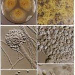
A Colonies on MEA +20 % sucrose after 2 weeks; B ascomata, x 40; C conidiophore of Aspergillus glaucus x 920;D conidiophore of Aspergillus glaucus x920 E. portion of ascoma with asci x 920. F ascospores x2330.
-
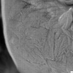
Scanning electron micrographs of A. fumigatus conidia of transformants rodB-02 (b). Size bar, 100 nm.

-
Scanning electron micrographs of A. fumigatus conidia of the wild-type G10 strain (a). Size bar, 100 nm.
-
Scanning electron micrographs of A. fumigatus conidia of rodA rodB-26 (d).Size bar, 100 nm.


 ,
, 
