Date: 26 November 2013
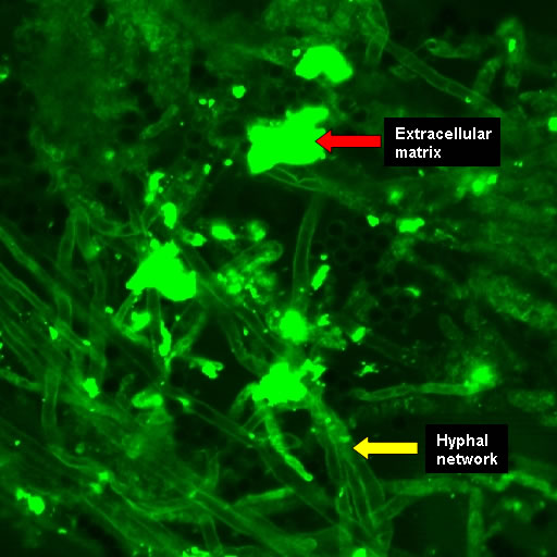
Biofilms on bronchial epithelial cells in vitro.
Copyright:
Images kindly donated by Frank-Michael C. Müller, Pediatric Pulmonology, Cystic Fibrosis Centre and Infectious Diseases, Department of Pediatrics III, University of Heidelberg, Im Neuenheimer Feld 430, D-69120 Heidelberg, Germany.
Notes:
Confocal scanning laser microscopy – (CLSM). CAAF stained showing the hyphal network in contact with extracellular matrix. The polysaccharides of the ECM and fungal cell walls were stained by CAAF (concanavalin A-Alexa Fluor 488 conjugate).
Images library
-
Title
Legend
-
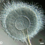
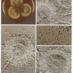
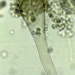
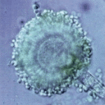
Aspergillus flavus Link. A colonies after 1 weekB,C conidial heads x 920D Conidia x920 E Conidial head x920
-
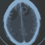
Patient diagnosed with Stage C Chronic Lymphocytic Leukaemia, treated in the MRC CLL4 study, with prednisolone for 4 weeks followed by oral chlorambucil for 7 days. Patient developed severe pneumonia due to pseudomonas and staphylococcus. Following treatment with broad spectrum antibiotics and 1 week of Abelcet, patient was readmitted with headache, disorientation and fever. CT brain scans showed 3 ring enhancing lesions, aspirated material showed neutrophils but grew aspergillus. Patient now improved on Abelcet (4 mg/kg) and oral itrconazole suspension 200mg b.d.
Thanks to Richard Chasty, Consultant Haematologist, North Staffordsire Hospital, UK.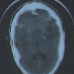 ,
, 

![Asp[1]niger Asp[1]niger](https://www.aspergillus.org.uk/wp-content/uploads/2013/11/Asp1niger-150x150.gif)


