Date: 26 November 2013
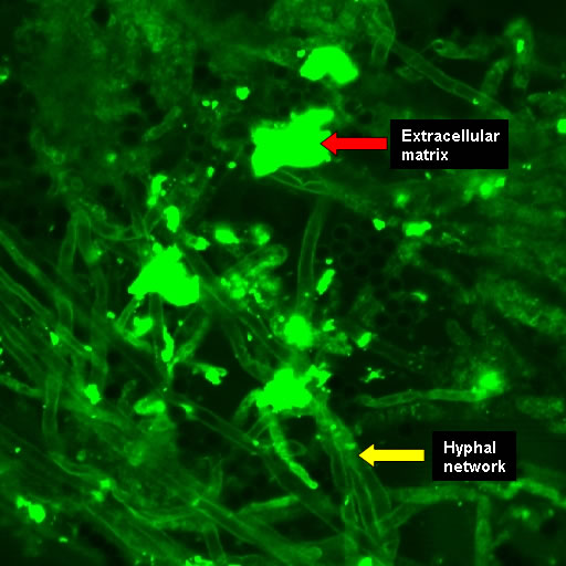
Biofilms on bronchial epithelial cells in vitro.
Copyright:
Images kindly donated by Frank-Michael C. Müller, Pediatric Pulmonology, Cystic Fibrosis Centre and Infectious Diseases, Department of Pediatrics III, University of Heidelberg, Im Neuenheimer Feld 430, D-69120 Heidelberg, Germany.
Notes:
Confocal scanning laser microscopy – (CLSM). CAAF stained showing the hyphal network in contact with extracellular matrix. The polysaccharides of the ECM and fungal cell walls were stained by CAAF (concanavalin A-Alexa Fluor 488 conjugate).
Images library
-
Title
Legend
-
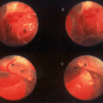
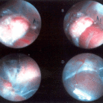
Falcons: The following images were obtained by endoscopy of falcons with aspergillosis.A,B Thoracic airsac (T) with prominent blood vessels and a dead serratospiculum worm (W). The presence of these lung worms makes the airsac look milky. D Normal ovary with developing follicles.

-
Falcons: The following images were obtained by endoscopy of falcons with aspergillosis.B,D Aspergillus lesions (A) over a swollen liver

-
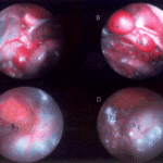
Falcons: The following images were obtained by endoscopy of falcons with aspergillosis.B Cranial, middle, caudal lobes (K1,K2,K3) of the left kidney, all the lobes show slight nephromegaly.C Yellow aspergillus colony (A1), lying adjacent to the lung.D White aspergillus colonies (A2,A3,A4).
-
Falcons: The following images were obtained by endoscopy of falcons with aspergillosis.C Cranial pole of left kidney (K) -mildly inflamed.D Ovary ( F) with developing follicles.
-
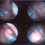
The following images were obtained by endoscopy of falcons with aspergillosis.A and B Lung Worm (S) over liver (Li) (serratospiculum seurati)C and D Aspergilloma (A) and prominent blood vessels on the caudal thoracic air sac (T).
