Date: 26 November 2013
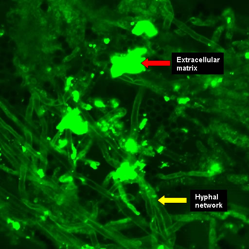
Biofilms on bronchial epithelial cells in vitro.
Copyright:
Images kindly donated by Frank-Michael C. Müller, Pediatric Pulmonology, Cystic Fibrosis Centre and Infectious Diseases, Department of Pediatrics III, University of Heidelberg, Im Neuenheimer Feld 430, D-69120 Heidelberg, Germany.
Notes:
Confocal scanning laser microscopy – (CLSM). CAAF stained showing the hyphal network in contact with extracellular matrix. The polysaccharides of the ECM and fungal cell walls were stained by CAAF (concanavalin A-Alexa Fluor 488 conjugate).
Images library
-
Title
Legend
-
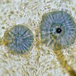
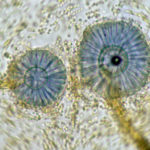
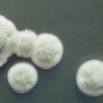
Scanning electron micrograph of Aspergillus ochraceopetaliformis conidial heads
-
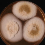
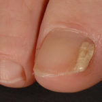
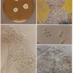
Image D & E. A case of onychomycosis associated with Aspergillus ochraceopetaliformis as described in Nail infection by Aspergillus ochraceopetaliformis. Med Mycol. 2009 Mar 9:1-5, 2009, Brasch J, Varga J, Jensen JM, Egberts F & Tintelnot K
 ,
,  ,
,  ,
,  ,
, 
-
Further details
Image 5. Oral itraconazole pulse therapy was given to the patient (200 mg twice daily for 1 week, with 3 weeks off between successive pulses, for four pulses) and treatment was successful.
 ,
,  ,
,  ,
,  ,
, 
-
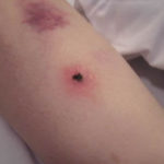
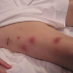
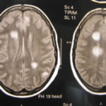
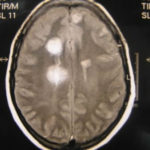
This patient was 28 yr old with adult lymphocytic leukaemia. She received induction chemotherapy and this infection developed 2 days after recovering from neutropenia.
 ,
,  ,
,  ,
,  ,
,  ,
,  ,
,  ,
,  ,
,  ,
, 
-
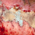
Close-up image of the lesion on the left thigh showing a mat of hyphae over the wound.
-
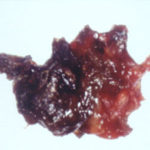
Eosinophilic mucin with A. flavus in the nasal cavity. Irregular crust of 2.5 cm from a patient diagnosed as allergic fungal sinusitis. Patient with allergic fungal sinusitis
-
GMS stain of eosinophilic mucin reveals a darkly stained dichotomously branched A. flavus hyphae within cellular background. Patient with allergic fungal sinusitis
