Date: 26 November 2013
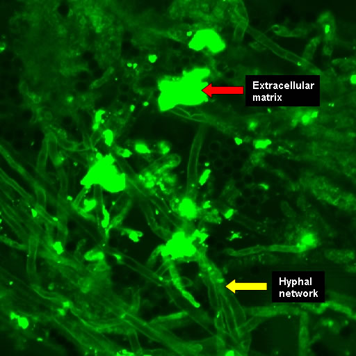
Biofilms on bronchial epithelial cells in vitro.
Copyright:
Images kindly donated by Frank-Michael C. Müller, Pediatric Pulmonology, Cystic Fibrosis Centre and Infectious Diseases, Department of Pediatrics III, University of Heidelberg, Im Neuenheimer Feld 430, D-69120 Heidelberg, Germany.
Notes:
Confocal scanning laser microscopy – (CLSM). CAAF stained showing the hyphal network in contact with extracellular matrix. The polysaccharides of the ECM and fungal cell walls were stained by CAAF (concanavalin A-Alexa Fluor 488 conjugate).
Images library
-
Title
Legend
-
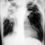
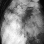
Right apical aspergilloma, patient WC. Plain chest radiograph of patient with right apical aspergilloma in an old, large tuberculous cavity. Severe haemoptysis and respiratory insufficiency together constituted the indications for embolisation which was done in one session over a 3 hour period (see images 1-6).
-
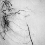
Embolisation 7 – patient WC. Angiogram of the lateral thoracic artery on subtraction film showing grossly abnormal vasculature inferiorly shunting along several anterior intercostal arteries to the internal mammary artery. In addition a pseudoaneurysm is shown.
-
Embolisation 6 – patient WC. Catheter tip in the lateral thoracic artery on screening film.
-
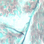
Grocott (silver) stain showing branching septate hyphae fairly typical of Aspergillus in mucus. The apparent right angle branching is unusual.
-
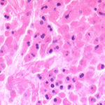
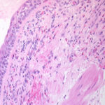
Bronchial mucosa under H & E stain showing numerous eosinophils deep to the mucosa, and mucus in the lumen of the bronchiole.
-
Grocott (silver) stain showing branching septate hyphae fairly typical of Aspergillus in mucus. The apparent right angle branching is unusual.

-
Severe kyphoscoliosis caused by greater than 40 years of prednisolone for ABPA and asthma.


