Date: 26 November 2013
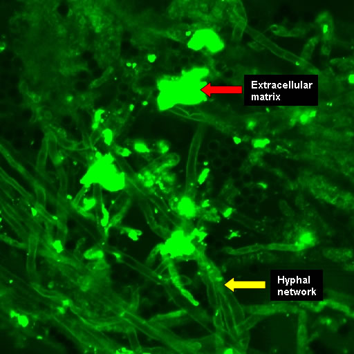
Biofilms on bronchial epithelial cells in vitro.
Copyright:
Images kindly donated by Frank-Michael C. Müller, Pediatric Pulmonology, Cystic Fibrosis Centre and Infectious Diseases, Department of Pediatrics III, University of Heidelberg, Im Neuenheimer Feld 430, D-69120 Heidelberg, Germany.
Notes:
Confocal scanning laser microscopy – (CLSM). CAAF stained showing the hyphal network in contact with extracellular matrix. The polysaccharides of the ECM and fungal cell walls were stained by CAAF (concanavalin A-Alexa Fluor 488 conjugate).
Images library
-
Title
Legend
-
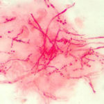
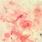
Corneal ulcer – gram stain. Corneal scrapings were taken from a 67 yr old farmer presenting with a corneal ulcer of the right eye. A piece of vegetable matter was embedded in the cornea and scrapings were done. Gram stain (500x magnification) showed numerous septate hyphae. Cultures grew a small amount of A fumigatus.

-
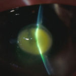
Aspergillus keratitis. Central lesion in aspergillus keratitis following a corneal foreign body which made a good response to topical treatment alone, albeit over 2 months intensive treatment.
-
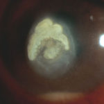
Aspergillus keratitis. B- Severe central aspergillus infection with a “cheesey†looking area of the lesion and hypopyon (fluid level of inflammatory cells in the anterior chamber)
-
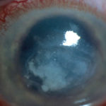
Aspergillus keratitis. A- Severe aspergillus infection with large area of corneal ulceration and deep stromal involvement
-
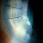
Candida keratitis. Focal candida keratitis as an unusual cause of a suture related infection following corneal transplantation for non infective indication
-
Candida keratitis. Subacute onset of candida keratitis in a young adult in whom dust blew into her eye in Greece. A slightly “feathery†edge to stromal involvement is suggestive of fungal infection
-
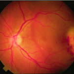
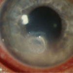
Aspergillus endopthalmitis. Temporal necrosis due to Aspergillus endopthalmitis as part of disseminated disease. No evidence of vitritis. Systemic treatment essential.
