Date: 7 May 2013
Copyright: n/a
Notes:
Colonies on CYA 40-60 mm diam, plane or lightly wrinkled, low, dense and velutinous or with a sparse, floccose overgrowth; mycelium inconspicuous, white; conidial heads borne in a continuous, densely packed layer, Greyish Turquoise to Dark Turquoise (24-25E-F5); clear exudate sometimes produced in small amounts; reverse pale or greenish. Colonies on MEA 40-60 mm diam, similar to those on CYA but less dense and with conidia in duller colours (24-25E-F3); reverse uncoloured or greyish. Colonies on G25N less than 10 mm diam, sometimes only germination, of white mycelium. No growth at 5°C. At 37°C, colonies covering the available area, i.e. a whole Petri dish in 2 days from a single point inoculum, of similar appearance to those on CYA at 25°C, but with conidial columns longer and conidia darker, greenish grey to pure grey.
Conidiophores borne from surface hyphae, stipes 200-400 µm long, sometimes sinuous, with colourless, thin, smooth walls, enlarging gradually into pyriform vesicles; vesicles 20-30 µm diam, fertile over half or more of the enlarged area, bearing phialides only, the lateral ones characteristically bent so that the tips are approximately parallel to the stipe axis; phialides crowded, 6-8 µm long; conidia spherical to subspheroidal, 2.5-3.0 µm diam, with finely roughened or spinose walls, forming radiate heads at first, then well defined columns of conidia.
Distinctive features
This distinctive species can be recognised in the unopened Petri dish by its broad, velutinous, bluish colonies bearing characteristic, well defined columns of conidia. Growth at 37°C is exceptionally rapid. Conidial heads are also diagnostic: pyriform vesicles bear crowded phialides which bend to be roughly parallel to the stipe axis. Care should be exercised in handling cultures of this species.
Images library
-
Title
Legend
-
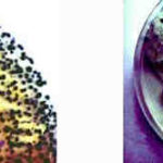
Left= an agar air plate exposed for 2 minutes after the barley had been turned. showing numerous colonies of the fungus following incubation at 26C on 2% malt agar.Right= A sputum sample taken from a maltworker after exposure showing many fungal colonies when cultured on agar. His commensal yeast flora is seen towards the right base as cream/white colonies.

-
growing on contaminated barley malt. The deep blue-green heads made up of chains of conidia are seen on the left. On the right, conidiophores from which conidial chains are developed show typical clavate heads. Stain- Cotton blue in Lactophenol.

-
Sections of colonised alveoli
On the left, the conidiophores sporulate freely, on the right they are seen to cease development at the phialide stage. Carbon deposits are clearly seen here.A=alveolus, As=alveolar septum, C=conidiophore, P=phialide
-
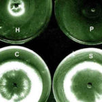
The growth isolated from the aspergilloma in the presence of living cells of the three bacterial species in culture.The most marked inhibition occurred with Pseudomonas aeruginosa(P) and Haemophilus influenzae(H) and to a much lesser extent with Staphylococcus aureus(S). C=control. Inhibitory factors were components of the bacterial slime layers.

-
aspergilloma removed from the lung cavity seen in section.Staining was with methenamine/silver and light green. The structure shows zonation probably due to variations in the growth rate of mycelium(deep brown). A mucus layer(stained green) containing cell debris and bacteria is seen shrouding the fungus. Bacterial genera were Staphylococcus, Haemophilus and Pseudodomonas.

-
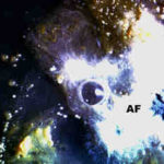
Plugs cultured on agarA young colony of the fungus(AF) has a central patch of sporulation and is surrounded by colonies of bacteria and yeasts.

-
A part of the mycelium in a stained sputum plug showing the dichotomously branched hyphae of Aspergillus fumigatus.


![Asp[1]fumighead2copyr Asp[1]fumighead2copyr](https://www.aspergillus.org.uk/wp-content/uploads/2013/11/Asp1fumighead2copyr.jpg)


