Date: 7 May 2013
Copyright: n/a
Notes:
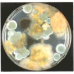
Colonies on CYA 40-60 mm diam, plane or lightly wrinkled, low, dense and velutinous or with a sparse, floccose overgrowth; mycelium inconspicuous, white; conidial heads borne in a continuous, densely packed layer, Greyish Turquoise to Dark Turquoise (24-25E-F5); clear exudate sometimes produced in small amounts; reverse pale or greenish. Colonies on MEA 40-60 mm diam, similar to those on CYA but less dense and with conidia in duller colours (24-25E-F3); reverse uncoloured or greyish. Colonies on G25N less than 10 mm diam, sometimes only germination, of white mycelium. No growth at 5°C. At 37°C, colonies covering the available area, i.e. a whole Petri dish in 2 days from a single point inoculum, of similar appearance to those on CYA at 25°C, but with conidial columns longer and conidia darker, greenish grey to pure grey.
Conidiophores borne from surface hyphae, stipes 200-400 µm long, sometimes sinuous, with colourless, thin, smooth walls, enlarging gradually into pyriform vesicles; vesicles 20-30 µm diam, fertile over half or more of the enlarged area, bearing phialides only, the lateral ones characteristically bent so that the tips are approximately parallel to the stipe axis; phialides crowded, 6-8 µm long; conidia spherical to subspheroidal, 2.5-3.0 µm diam, with finely roughened or spinose walls, forming radiate heads at first, then well defined columns of conidia.
Distinctive features
This distinctive species can be recognised in the unopened Petri dish by its broad, velutinous, bluish colonies bearing characteristic, well defined columns of conidia. Growth at 37°C is exceptionally rapid. Conidial heads are also diagnostic: pyriform vesicles bear crowded phialides which bend to be roughly parallel to the stipe axis. Care should be exercised in handling cultures of this species.
Images library
-
Title
Legend
-
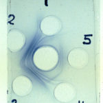
Aspergillus – Immunodiffusion Test showing stong immunoprecipitin lines against aspergillus.
-
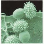
Talaromyces macrosporus- very short heat treatments of dormant ascospores at 85 C are able to activate these spores to germinate within 1 minute. One stage of germination is a very quick bursting of the thinly walled inner cell through the thick ornamented outer cell wall of the spore.
-
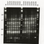
RAPD profiles of 14 A fumigatus isolates from Swedish and New Zealand saw mills, generated with primer R108. Mwt markers (lambda with Pst1) indicated by M.
-
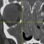
These 3 images show non-union of the sternum post-aortic valve replacement as a result of local Aspergillus fumigatus infection. Underlying the sternum is some soft tissue which is presumptively also infection and in the most inferior image, the pericardium is also invoved.
The patient also is diabetic, with rhematoid arthritis and ulcerative colitis and occasionally receives a course of corticosteroids. The wound discharged pus which grew A. fumigatus. It was managed conservatively with itraconazole initally, with failure of therapy over 3 months.
 ,
,  ,
, 
-
CHEF gel of A. fumigatus chromosomes. The electrophoretic karyotype of two isolates of A. fumigatus is shown. The chromosomal bands have been resolved using Bio-Rad CHEF DRII equipment. The sizes of the bands vary between the two isolates and at least five bands have been resolved using the following conditions: chromosomal grade agarose (Bio-Rad) at 1.2 % in 1 x TAE buffer was used; the gel was run with a field strength of 1.8 V/cm for 48 h with a switch time of 2200 s and for 71 h with a switching ramp

-
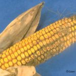
Aspergillus ear rot and storage mould – Aspergillus flavus and Aspergillus parasiticus can produce aflatoxins are generally known as storage fungi, but they can also cause ear rots in the field. These species are observed as a gray-green, powdery molds and they can be detected in corn because they produce compounds that are fluorescent under black light.
-
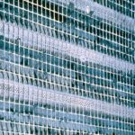
Environmental and sick building images. Dirty ventilation intake ducts

![Asp[1]fumighead2copyr Asp[1]fumighead2copyr](https://www.aspergillus.org.uk/wp-content/uploads/2013/11/Asp1fumighead2copyr.jpg)
