Date: 7 May 2013
Copyright: n/a
Notes:
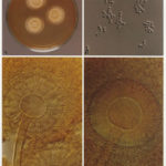
Colonies on CYA 40-60 mm diam, plane or lightly wrinkled, low, dense and velutinous or with a sparse, floccose overgrowth; mycelium inconspicuous, white; conidial heads borne in a continuous, densely packed layer, Greyish Turquoise to Dark Turquoise (24-25E-F5); clear exudate sometimes produced in small amounts; reverse pale or greenish. Colonies on MEA 40-60 mm diam, similar to those on CYA but less dense and with conidia in duller colours (24-25E-F3); reverse uncoloured or greyish. Colonies on G25N less than 10 mm diam, sometimes only germination, of white mycelium. No growth at 5°C. At 37°C, colonies covering the available area, i.e. a whole Petri dish in 2 days from a single point inoculum, of similar appearance to those on CYA at 25°C, but with conidial columns longer and conidia darker, greenish grey to pure grey.
Conidiophores borne from surface hyphae, stipes 200-400 µm long, sometimes sinuous, with colourless, thin, smooth walls, enlarging gradually into pyriform vesicles; vesicles 20-30 µm diam, fertile over half or more of the enlarged area, bearing phialides only, the lateral ones characteristically bent so that the tips are approximately parallel to the stipe axis; phialides crowded, 6-8 µm long; conidia spherical to subspheroidal, 2.5-3.0 µm diam, with finely roughened or spinose walls, forming radiate heads at first, then well defined columns of conidia.
Distinctive features
This distinctive species can be recognised in the unopened Petri dish by its broad, velutinous, bluish colonies bearing characteristic, well defined columns of conidia. Growth at 37°C is exceptionally rapid. Conidial heads are also diagnostic: pyriform vesicles bear crowded phialides which bend to be roughly parallel to the stipe axis. Care should be exercised in handling cultures of this species.
Images library
-
Title
Legend
-
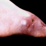
This photo shows extensive infection of the burn wound on the leg of a 7 year old boy, acquired about 3 weeks after the injury. Despite medical therapy he died.

-
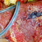
This patient, had had a laparostomy for recurrent intra-abdominal sepsis following on from crohns disease. She was transferred to another intensive care unit and her dressings changed daily. Shortly after, this dark patches appeared on her liver (as seen here A) and her colon. Superficial biopsies and culture showed A.fumigatus invading liver capsule. She responded to amphotericin B therapy.
B shows patient after treatment.
 ,
, 
-
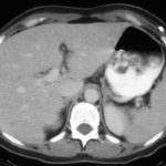
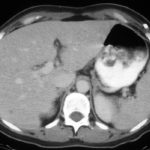
Hepatic aspergillosis, pt KO. Repeat CT scan of the liver showing almost complete resolution of lesions on itraconazole therapy.
-
Image A. The CT scan of her abdomen had the appearances shown here. She also has small pulmonary nodules. Bioposy of the liver revealed hyphae consistent with Aspergillus.
Image B. She responded well to oral itraconazole therapy.
 ,
, 
-
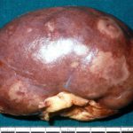
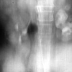
This image shows the pelvis of the left kidney filled with fungal balls. Eventually, after failing amphotericin B therapy, she required a nephrectomy. Her case is reported in Davies SP, Webb WJS, Patou G, Murray WK, Denning DW. Renal aspergilloma – a case illustrating the problems of medical therapy. Nephrol Dial Transplant 1987; 2: 568-572.
-
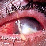
Aspergillus keratitis. Good example of Aspergillus keratitis caused by A.glaucus. Usually A.fumitagus and A.flavus are the causes.

![Asp[1]fumighead2copyr Asp[1]fumighead2copyr](https://www.aspergillus.org.uk/wp-content/uploads/2013/11/Asp1fumighead2copyr.jpg)
 ,
, 