Date: 7 May 2013
Copyright: n/a
Notes:
Colonies on CYA 40-60 mm diam, plane or lightly wrinkled, low, dense and velutinous or with a sparse, floccose overgrowth; mycelium inconspicuous, white; conidial heads borne in a continuous, densely packed layer, Greyish Turquoise to Dark Turquoise (24-25E-F5); clear exudate sometimes produced in small amounts; reverse pale or greenish. Colonies on MEA 40-60 mm diam, similar to those on CYA but less dense and with conidia in duller colours (24-25E-F3); reverse uncoloured or greyish. Colonies on G25N less than 10 mm diam, sometimes only germination, of white mycelium. No growth at 5°C. At 37°C, colonies covering the available area, i.e. a whole Petri dish in 2 days from a single point inoculum, of similar appearance to those on CYA at 25°C, but with conidial columns longer and conidia darker, greenish grey to pure grey.
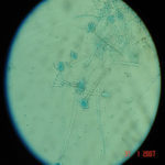
Conidiophores borne from surface hyphae, stipes 200-400 µm long, sometimes sinuous, with colourless, thin, smooth walls, enlarging gradually into pyriform vesicles; vesicles 20-30 µm diam, fertile over half or more of the enlarged area, bearing phialides only, the lateral ones characteristically bent so that the tips are approximately parallel to the stipe axis; phialides crowded, 6-8 µm long; conidia spherical to subspheroidal, 2.5-3.0 µm diam, with finely roughened or spinose walls, forming radiate heads at first, then well defined columns of conidia.
Distinctive features
This distinctive species can be recognised in the unopened Petri dish by its broad, velutinous, bluish colonies bearing characteristic, well defined columns of conidia. Growth at 37°C is exceptionally rapid. Conidial heads are also diagnostic: pyriform vesicles bear crowded phialides which bend to be roughly parallel to the stipe axis. Care should be exercised in handling cultures of this species.
Images library
-
Title
Legend
-
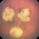
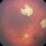
Bilateral A. fumigatus endophthalmitis in association with pulmonary and cerebral aspergillosis, complicating severe autoimmune disease treated with intense immunosuppression. Right eye.
-
Bilateral A. fumigatus endophthalmitis in association with pulmonary and cerebral aspergillosis, complicating severe autoimmune disease treated with intense immunosuppression. Left eye.
-
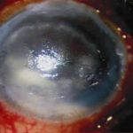
Aspergillus keratitis – a case of fungal keratitis following amnoiotic membrane transplantation (AMT) for bullous keratopathy. Slit-lamp photograph of left eye showing ring shaped stromal infiltrate.
-
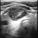
A 14 year old boy with a T cell lymphoma received induction chemotherapy with high dose dexamethasone. At no time during this therapy was he neutropenic. Three weeks into treatment his dexamethasone was reduced and stopped due to gastro-intestinal side effects. On recovery of his abdominal symptoms, he developed sudden onset of severe right sided pleuritic chest pain. Initially, he was thought to have a pulmonary embolism as a chest X-ray showed a solid wedge shaped area of consolidation. He then developed a cough and temperature and sputum grew Aspergillus fumigatus. The wedge shaped lesion developed cavitation. Despite AmBisome at 5mg/Kg, commenced within 4 days of onset of symptoms, the chest X-ray appearance got gradually worse over the following week. This led to substitution of Ambisome by caspofungin and voriconazole. He developed a thyroid cyst with haemorrhage that on aspiration grew A. fumigatus. His chest X-ray continued to worsen. He underwent a right lower lobe lobectomy, which confirmed the diagnosis of invasive pulmonary aspergillosis. Unfortunately the thyroid cyst continued to increase in size resulting in removal of the right lobe of the thyroid.
Chest CT scan carried out 3 weeks after lobectomy revealed new lesions within the right upper lobe of the lung. At this time the voriconazole therapy was stopped and posaconazole started. The area of the thyroid remains free of Aspergillus and the lung lesions appear to be improving 6 weeks after the start of posaconazole.
 ,
,  ,
, 
-
Aspergillus keratitis. Severe peripheral lesion due to aspergillus unlikely to respond well to treatment.

-
Aspergillus keratitis. Corneal scar at end of treatment of case in previous slide.Vision recovered to 6/9.

-
Aspergillus keratitis. Severe central aspergillus infection with a “cheesey†looking area of the lesion and hypopyon (fluid level of inflammatory cells in the anterior chamber)


![Asp[1]fumighead2copyr Asp[1]fumighead2copyr](https://www.aspergillus.org.uk/wp-content/uploads/2013/11/Asp1fumighead2copyr.jpg)
