Date: 7 May 2013
Copyright: n/a
Notes:
Colonies on CYA 40-60 mm diam, plane or lightly wrinkled, low, dense and velutinous or with a sparse, floccose overgrowth; mycelium inconspicuous, white; conidial heads borne in a continuous, densely packed layer, Greyish Turquoise to Dark Turquoise (24-25E-F5); clear exudate sometimes produced in small amounts; reverse pale or greenish. Colonies on MEA 40-60 mm diam, similar to those on CYA but less dense and with conidia in duller colours (24-25E-F3); reverse uncoloured or greyish. Colonies on G25N less than 10 mm diam, sometimes only germination, of white mycelium. No growth at 5°C. At 37°C, colonies covering the available area, i.e. a whole Petri dish in 2 days from a single point inoculum, of similar appearance to those on CYA at 25°C, but with conidial columns longer and conidia darker, greenish grey to pure grey.
Conidiophores borne from surface hyphae, stipes 200-400 µm long, sometimes sinuous, with colourless, thin, smooth walls, enlarging gradually into pyriform vesicles; vesicles 20-30 µm diam, fertile over half or more of the enlarged area, bearing phialides only, the lateral ones characteristically bent so that the tips are approximately parallel to the stipe axis; phialides crowded, 6-8 µm long; conidia spherical to subspheroidal, 2.5-3.0 µm diam, with finely roughened or spinose walls, forming radiate heads at first, then well defined columns of conidia.
Distinctive features
This distinctive species can be recognised in the unopened Petri dish by its broad, velutinous, bluish colonies bearing characteristic, well defined columns of conidia. Growth at 37°C is exceptionally rapid. Conidial heads are also diagnostic: pyriform vesicles bear crowded phialides which bend to be roughly parallel to the stipe axis. Care should be exercised in handling cultures of this species.
Images library
-
Title
Legend
-
Further details
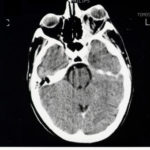
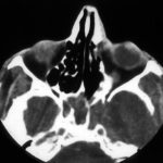
Image A. The bone appears normal, but the features are most consistent with infection, less so with a pseudotumour. Bone windows not shown. Biopsy demonstrated hyphal invasion and cultures grew A. fumigatus.
 ,
,  ,
,  ,
, 
-
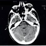
Bilateral Aspergillus osteomyelitis of the base of the skull in a non-immunocompromised woman, aged 38, following Aspergillus sinusitis. This was eventually cured with an 18 month course of voriconazole.
-
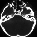
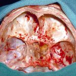
In this non-immunocompromised patient with histologically and culture proven invasive aspergillosis most of the landmarks have been surgically removed during 3 operations.

-
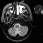
The maxillary sinus on the left is completely opacified with high signal, The high signal seen in the maxillary sinus on the right is of marginal significance. The sinuses and other cerebellum are normal.
-
CT scan of the sinuses shows a completely opacified sphenoid sinus in this man with AIDS (Khoo S, Denning DW. Invasive aspergillosis in patients with AIDS Clin Infect Dis 1994; 19 (suppl 1): S41-8.) He had complained of headache for about 3 months before this scan was done. In addition to sinus disease, erosion of disease through the left lateral wall of the sinus into the arterior horn of the temporal lobe, can be seen leading to a cerebral abscess. The patient was treated with itraconazole, but had undetectable serum concentrations because of prior phenytoin, and died after nine days of therapy of an MRSA bacteraemia. He was reported as an “other failure” in Denning DW, Lee JY, Hostetler JS, Pappas P, Kauffman CA, Dewsnup DH, Galgiani JN, Graybill JR, Sugar AM, Catanzaro A, Gallis H, Perfect JR, Dockery B, Dismukes WE, Stevens DA, NIAID Mycoses Study Group multicenter trial of oral itraconazole therapy of invasive aspergillosis. Am J Med 1994; 97: 135-144.
Cultures from the sinus and brain abcess grew A.fumigatus.
 ,
, 

![Asp[1]fumighead2copyr Asp[1]fumighead2copyr](https://www.aspergillus.org.uk/wp-content/uploads/2013/11/Asp1fumighead2copyr.jpg)


