Date: 7 May 2013
Copyright: n/a
Notes:
Colonies on CYA 40-60 mm diam, plane or lightly wrinkled, low, dense and velutinous or with a sparse, floccose overgrowth; mycelium inconspicuous, white; conidial heads borne in a continuous, densely packed layer, Greyish Turquoise to Dark Turquoise (24-25E-F5); clear exudate sometimes produced in small amounts; reverse pale or greenish. Colonies on MEA 40-60 mm diam, similar to those on CYA but less dense and with conidia in duller colours (24-25E-F3); reverse uncoloured or greyish. Colonies on G25N less than 10 mm diam, sometimes only germination, of white mycelium. No growth at 5°C. At 37°C, colonies covering the available area, i.e. a whole Petri dish in 2 days from a single point inoculum, of similar appearance to those on CYA at 25°C, but with conidial columns longer and conidia darker, greenish grey to pure grey.
Conidiophores borne from surface hyphae, stipes 200-400 µm long, sometimes sinuous, with colourless, thin, smooth walls, enlarging gradually into pyriform vesicles; vesicles 20-30 µm diam, fertile over half or more of the enlarged area, bearing phialides only, the lateral ones characteristically bent so that the tips are approximately parallel to the stipe axis; phialides crowded, 6-8 µm long; conidia spherical to subspheroidal, 2.5-3.0 µm diam, with finely roughened or spinose walls, forming radiate heads at first, then well defined columns of conidia.
Distinctive features
This distinctive species can be recognised in the unopened Petri dish by its broad, velutinous, bluish colonies bearing characteristic, well defined columns of conidia. Growth at 37°C is exceptionally rapid. Conidial heads are also diagnostic: pyriform vesicles bear crowded phialides which bend to be roughly parallel to the stipe axis. Care should be exercised in handling cultures of this species.
Images library
-
Title
Legend
-
Pulmonary aspergillosis (Grocott) (cow 2). Serial section of the lesion previously described for cow 2 stained by Grocott. Aspergillus spp. and Actinomyces pyogenes were cultured from the lesions.

-
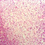
Pulmonary aspergillosis (cow 2). Taken from the edge of a lesion, as described in the previous image for cow 2.
-
Pulmonary aspergillosis (cow 2). Section of lung from a 2 year old cow with weight loss and anorexia since calving. On necropsy examination, multiple firm masses were identified throughout the lungs. These were cavitating in nature, with a necrotic centre and peripheral fibrosis. Both this section and the following one are taken from the edge of such a lesion and demonstrate the pyogranulomatous inflammatory response.
-
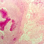
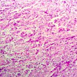
Guttural pouch aspergillus (PAS) (horse TW). Serial section of previous slide for horse TW stained by PAS reveals the presence of fungal hyphae within the inflammatory tissue. Aspergillus spp. is the likely cause of equine guttural pouch mycosis.
-
Guttural pouch aspergillosis (horse TW). Section of the wall of the guttural pouch from an 8 year old, neutered male horse with recurrent right-sided epistaxis of 10 days duration. Endoscopic examination confirmed the origin of the blood to be the guttural pouch and the right internal carotid artery was ligated surgically. Necropsy examination revealed an extensive ruptured aneurysm of this vessel and guttural pouch mycosis. The section reveals necrosis and chronic inflammatory infiltration of guttural pouc
-
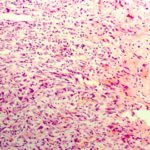
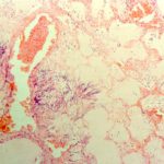
Pulmonary aspergillosis. Section of lung from a 6 year old, Holstein cow with mycotic pneumonia attributed to Aspergillus spp.
-
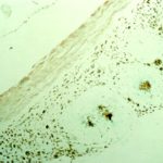
Meningitis (dog R). Grocott-stained serial section of the previous slide for dog R reveals the presence of fungal material at the centre of each microgranuloma. These lesions were not cultured, but Aspergillus spp. was identified by immunohistochemical examination using a panel of specific antisera. There was no evidence of systemic involvement in this dog.
-
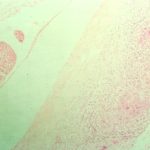
Aspergillus meningitis (dog R). Section of spinal meninges from a 4 year old, neutered female German shepherd dog with multiple discospondylitis involving T1-2, T6-7, T10-11 and L5-6. There are a series of coalescing microgranulomas associated with meninges at each site.
-
Pulmonary aspergillosis (cat T). Section of lung from a young adult, female domestic short haired cat. This was one of seven unvaccinated farm cats that died over a 12 month period. At necropsy examination there was diffuse pulmonary consolidation with scattered abscessation. Aspergillus spp. was cultured from these lesions. The cat also had histopathological evidence of lymph node atrophy, a change that may be attributed to feline immunodeficiency virus (FIV) infection.


![Asp[1]fumighead2copyr Asp[1]fumighead2copyr](https://www.aspergillus.org.uk/wp-content/uploads/2013/11/Asp1fumighead2copyr.jpg)
