Date: 26 November 2013
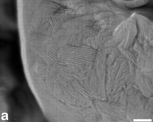
Scanning electron micrographs of A. fumigatus conidia of the wild-type G10 strain (a). Size bar, 100 nm.
Copyright:
© Fungal Research Trust
Notes:
Scanning electron micrograph of an A.fumigatus conidium showing the hydrophobic rodlets covering the surface. The surface of many fungal conidia is covered by a thin layer of regularly arranged rodlets. This structure appears to favor air buoyancy and dispersion of the conidia by air currents.See Ref: Paris et al APPLIED AND ENVIRONMENTAL MICROBIOLOGY, Mar. 2003, 1581–1588.
Colonies on CYA 40-60 mm diam, plane or lightly wrinkled, low, dense and velutinous or with a sparse, floccose overgrowth; mycelium inconspicuous, white; conidial heads borne in a continuous, densely packed layer, Greyish Turquoise to Dark Turquoise (24-25E-F5); clear exudate sometimes produced in small amounts; reverse pale or greenish. Colonies on MEA 40-60 mm diam, similar to those on CYA but less dense and with conidia in duller colours (24-25E-F3); reverse uncoloured or greyish. Colonies on G25N less than 10 mm diam, sometimes only germination, of white mycelium. No growth at 5°C. At 37°C, colonies covering the available area, i.e. a whole Petri dish in 2 days from a single point inoculum, of similar appearance to those on CYA at 25°C, but with conidial columns longer and conidia darker, greenish grey to pure grey.
Conidiophores borne from surface hyphae, stipes 200-400 µm long, sometimes sinuous, with colourless, thin, smooth walls, enlarging gradually into pyriform vesicles; vesicles 20-30 µm diam, fertile over half or more of the enlarged area, bearing phialides only, the lateral ones characteristically bent so that the tips are approximately parallel to the stipe axis; phialides crowded, 6-8 µm long; conidia spherical to subspheroidal, 2.5-3.0 µm diam, with finely roughened or spinose walls, forming radiate heads at first, then well defined columns of conidia.
Distinctive features
This distinctive species can be recognised in the unopened Petri dish by its broad, velutinous, bluish colonies bearing characteristic, well defined columns of conidia. Growth at 37°C is exceptionally rapid. Conidial heads are also diagnostic: pyriform vesicles bear crowded phialides which bend to be roughly parallel to the stipe axis. Care should be exercised in handling cultures of this species.
Images library
-
Title
Legend
-
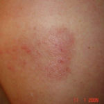
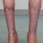
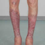
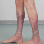
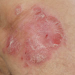
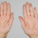
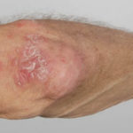
After 3 weeks of posaconazole given for chronic pulmonary aspergillosis, patient NC had a remarkable exacerbation of psoriasis. He had had psoriasis for years, with little trouble and almost no treatment. After taking posaconazole 400mg twice daily, he developed psoriatic plaques on his hands for the first time ever. The plaques on his lower legs became confluent. This occurred in association with worsening chest symptoms, notably increased coughing, more breathlessness and increasing oxygen requirement.
Posaconazole was stopped after 3 weeks, and 2 weeks later he was still very symptomatic with his chest. This responded to a 2 week course of corticosteroids, and his psoriasis also improved.
 ,
,  ,
,  ,
,  ,
,  ,
, 
-
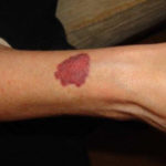
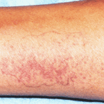
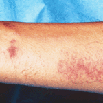
Patient PC: An example of localised caspofungin rash and phlebitis related to caspofungin infusion.
 ,
, 
-
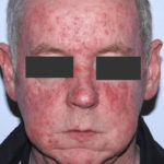
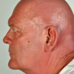
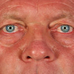
This 55 year old man with asthma, ABPA, severe bronchiectasis and lung fibrosis was treated with voriconazole, starting in June 2010. He had developed increasing dyspnoea on itraconazole for over 7 years, and his total IgE remained at 1100 KIU/L. He had marked photopsia (visual hallucinations) and facial erythema in the first 3 weeks of therapy. His trough voriconazole concentration was 1.17 mg/L. Over 3 months, he had minor improvement in his breathlessness but continued facial erythema, despite factor 50 sunblock. After 5 months of therapy his facial rash has altered to show acneiform lesions with localised crusting and background severe erythema. His face effectively crusted over, and he stopped therapy.
Over the next 3 weeks his facial appearance slowly improved .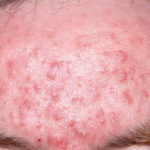 ,
,  ,
, 