Date: 26 November 2013
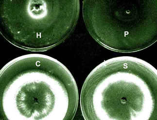
The growth isolated from the aspergilloma in the presence of living cells of the three bacterial species in culture.The most marked inhibition occurred with Pseudomonas aeruginosa(P) and Haemophilus influenzae(H) and to a much lesser extent with Staphylococcus aureus(S). C=control. Inhibitory factors were components of the bacterial slime layers.
Copyright:
Fungal Research Trust
Notes:
Colonies on CYA 40-60 mm diam, plane or lightly wrinkled, low, dense and velutinous or with a sparse, floccose overgrowth; mycelium inconspicuous, white; conidial heads borne in a continuous, densely packed layer, Greyish Turquoise to Dark Turquoise (24-25E-F5); clear exudate sometimes produced in small amounts; reverse pale or greenish. Colonies on MEA 40-60 mm diam, similar to those on CYA but less dense and with conidia in duller colours (24-25E-F3); reverse uncoloured or greyish. Colonies on G25N less than 10 mm diam, sometimes only germination, of white mycelium. No growth at 5°C. At 37°C, colonies covering the available area, i.e. a whole Petri dish in 2 days from a single point inoculum, of similar appearance to those on CYA at 25°C, but with conidial columns longer and conidia darker, greenish grey to pure grey.
Conidiophores borne from surface hyphae, stipes 200-400 µm long, sometimes sinuous, with colourless, thin, smooth walls, enlarging gradually into pyriform vesicles; vesicles 20-30 µm diam, fertile over half or more of the enlarged area, bearing phialides only, the lateral ones characteristically bent so that the tips are approximately parallel to the stipe axis; phialides crowded, 6-8 µm long; conidia spherical to subspheroidal, 2.5-3.0 µm diam, with finely roughened or spinose walls, forming radiate heads at first, then well defined columns of conidia.
Distinctive features
This distinctive species can be recognised in the unopened Petri dish by its broad, velutinous, bluish colonies bearing characteristic, well defined columns of conidia. Growth at 37°C is exceptionally rapid. Conidial heads are also diagnostic: pyriform vesicles bear crowded phialides which bend to be roughly parallel to the stipe axis. Care should be exercised in handling cultures of this species.
Images library
-
Title
Legend
-
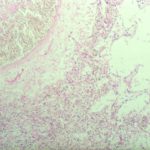
Pulmonary aspergillosis (PAS) (parrot C). Section of lung from parrot C stained by periodic acid schiff (PAS) demonstrating the hyphal material. Aspergillus spp and Bacillus cereus were cultured from the lesions.
-
Pulmonary aspergillosis (K&E) (parrot C). Tissue from an individually housed and recently purchased, 6 month old African grey parrot found dead in the cage. Necropsy examination revealed focal necrosis of the left lung. This section stained by haematoxylin and eosin reveals septate fungal hyphae within the lung parenchyma. Similar hyphae were located in the walls and lumen of parabronchi, and within the walls of pulmonary blood vessels.

-
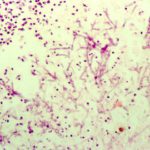
Nasal aspergillosis. Tissue from an 8 year old, neutered male thoroughbred horse with an initial history of sinusitis leading to progressive neurological signs (ataxia, behavioural abnormalities) and prolonged recumbency. Necropsy examination revealed a focus of grey-caseous material within the right nasal chamber that comprised a mat of branching, septae fungal hyphae and mixed inflammatory cells (haematoxylin and eosin stain). Aspergillus spp was cultured from the lesion. There was no gross or histologica
-
Immunofluorescence. Section of renal granuloma from dog J stained with polyclonal antiserum specific for Aspergillus terreus by immunofluorescence.

-
Complement deposition (dog J). Section of myocardial granuloma from dog J stained for canine complement C3 by immunofluorescence. Deposition of C3, but not C4, on fungal hyphae suggests activation of the alternative rather than classical pathway of complement.

-
Lymph node granuloma – Section of lymph node granuloma from a German shepherd dog with disseminated aspergillosis stained for canine IgA by immunofluorescence. The fungal hyphae within the centre of the lesion have surface IgA, and IgA-bearing plasma cells are present within the surrounding inflammatory infiltrate

-
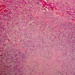
Extensive focus of pyogranulomatous inflammation within the kidney of dog J
-
Aspergillus granuloma within the myocardium of dog J.

-
Retinal aspergillosis (dog J) – Section of retina from a German shepherd dog with disseminated aspergillosis. Fungal hyphae and inflammatory cells are found within the vitreous


