Date: 26 November 2013
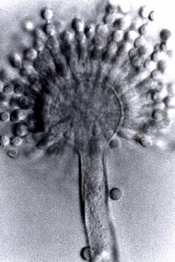
Aspergillus flavus
Copyright:
© Fungal Research Trust
Notes:
Colonies on CYA 60-70 mm diam, plane, sparse to moderately dense, velutinous in marginal areas at least, often floccose centrally, sometimes deeply so; mycelium only conspicuous in floccose areas, white; conidial heads usually borne uniformly over the whole colony, but sparse or absent in areas of floccose growth or sclerotial production, characteristically Greyish Green to Olive Yellow (1-2B-E5-7), but sometimes pure Yellow (2-3A7-8), becoming greenish in age; sclerotia produced by about 50% of isolates, at first white, becoming deep reddish brown, density varying from inconspicuous to dominating colony appearance and almost entirely suppressing conidial production; exudate sometimes produced, clear, or reddish brown near sclerotia; reverse uncoloured or brown to reddish brown beneath sclerotia. Colonies on MEA 50-70 mm diam, similar to those on CYA although usually less dense. Colonies on G25N 25-40 mm diam, similar to those on CYA or more deeply floccose and with little conidial production, reverse pale to orange or salmon. No growth at 5°C. At 37°C, colonies usually 55-65 mm diam, similar to those on CYA at 25°C, but more velutinous, with olive conidia, and sometimes with more abundant sclerotia.
Sclerotia produced by some isolates, at first white, rapidly becoming hard and reddish brown to black, spherical, usually 400- 800 µm diam. Teleomorph not known. Conidiophores borne from subsurface or surface hyphae, stipes 400 µm to 1 mm or more long, colourless or pale brown, rough walled; vesicles spherical, 20-45 µm diam, fertile over three quarters of the surface, typically bearing both metulae and phialides, but in some isolates a proportion or even a majority of heads with phialides alone; metulae and phialides of similar size, 7-10 µm long; conidia spherical to subspheroidal, usually 3.5-5.0 µm diam, with relatively thin walls, finely roughened or, rarely, smooth.
Distinctive features
Aspergillus flavus is distinguished by rapid growth at both 25°C and 37°C, and a bright yellow green (or less commonly yellow) conidial colour. A. flavus produces conidia which are rather variable in shape and size, have relatively thin walls, and range from smooth to moderately rough, the majority being finely rough.
Images library
-
Title
Legend
-
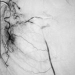
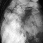
Embolisation 7 – patient WC. Angiogram of the lateral thoracic artery on subtraction film showing grossly abnormal vasculature inferiorly shunting along several anterior intercostal arteries to the internal mammary artery. In addition a pseudoaneurysm is shown.
-
Embolisation 6 – patient WC. Catheter tip in the lateral thoracic artery on screening film.
-
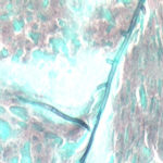
Grocott (silver) stain showing branching septate hyphae fairly typical of Aspergillus in mucus. The apparent right angle branching is unusual.
-
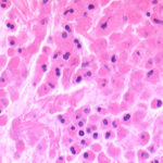
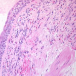
Bronchial mucosa under H & E stain showing numerous eosinophils deep to the mucosa, and mucus in the lumen of the bronchiole.
-
Grocott (silver) stain showing branching septate hyphae fairly typical of Aspergillus in mucus. The apparent right angle branching is unusual.

-
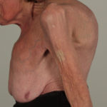
Severe kyphoscoliosis caused by greater than 40 years of prednisolone for ABPA and asthma.

-
These pictures show remarkable curvature of the spine as a result of collapse of the vertebral bodies of the thoracic vertebrae. This is a gross example of steroid-induced osteoporosis. The dose was not large in the last 10 years, typically 5-10mg daily, but multiple high dose courses and slow tapering lead to this outcome.
Her corticosteroid warning card is also demonstrated, as additional steroids are required for any significant illness or surgery, as her adrenal glands had completely atrophied.
Kindly supplied by Prof David Denning, South Manchester University Hospitals NHS Trust, Manchester UK
(© Fungal Research Trust)
 ,
,  ,
,  ,
,  ,
, 