Date: 26 November 2013
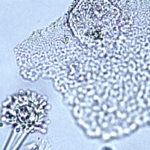
cultures grown from BAL fluid showing formation of sclerotia.
Copyright:
Kindly donated by Dr Claudia Venturelli and Dr Giorgia Bertazzoni, Laboratory of Microbiology – Policlinico of Modena-Italy. © Fungal Research Trust
Notes:
These colonies were isolated from a BAL, (also with bacterial qrowth of S.aureus and S.maltophilia) from a patient with a VAP (undergoing corticosteroid treatment). The growth medium used is sabouraud dextrose agar , incubated at 37° C The identification is made by microscopic/macroscopic observation criteria.
Colonies on CYA 60-70 mm diam, plane, sparse to moderately dense, velutinous in marginal areas at least, often floccose centrally, sometimes deeply so; mycelium only conspicuous in floccose areas, white; conidial heads usually borne uniformly over the whole colony, but sparse or absent in areas of floccose growth or sclerotial production, characteristically Greyish Green to Olive Yellow (1-2B-E5-7), but sometimes pure Yellow (2-3A7-8), becoming greenish in age; sclerotia produced by about 50% of isolates, at first white, becoming deep reddish brown, density varying from inconspicuous to dominating colony appearance and almost entirely suppressing conidial production; exudate sometimes produced, clear, or reddish brown near sclerotia; reverse uncoloured or brown to reddish brown beneath sclerotia. Colonies on MEA 50-70 mm diam, similar to those on CYA although usually less dense. Colonies on G25N 25-40 mm diam, similar to those on CYA or more deeply floccose and with little conidial production, reverse pale to orange or salmon. No growth at 5°C. At 37°C, colonies usually 55-65 mm diam, similar to those on CYA at 25°C, but more velutinous, with olive conidia, and sometimes with more abundant sclerotia.
Sclerotia produced by some isolates, at first white, rapidly becoming hard and reddish brown to black, spherical, usually 400- 800 µm diam. Teleomorph not known. Conidiophores borne from subsurface or surface hyphae, stipes 400 µm to 1 mm or more long, colourless or pale brown, rough walled; vesicles spherical, 20-45 µm diam, fertile over three quarters of the surface, typically bearing both metulae and phialides, but in some isolates a proportion or even a majority of heads with phialides alone; metulae and phialides of similar size, 7-10 µm long; conidia spherical to subspheroidal, usually 3.5-5.0 µm diam, with relatively thin walls, finely roughened or, rarely, smooth.
Distinctive features
Aspergillus flavus is distinguished by rapid growth at both 25°C and 37°C, and a bright yellow green (or less commonly yellow) conidial colour. A. flavus produces conidia which are rather variable in shape and size, have relatively thin walls, and range from smooth to moderately rough, the majority being finely rough.
Images library
-
Title
Legend
-
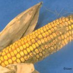
Aspergillus flavus and Aspergillus parasiticus can produce aflatoxins are generally known as storage fungi, but they can also cause ear rots in the field. These species are observed as a gray-green, powdery molds and they can be detected in corn because they produce compounds that are fluorescent under black light.
-
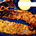
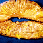
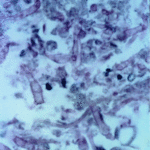
Histopathology of the jejenum showing necrosis and hyphae consistent with Aspergillus
-
Further image details
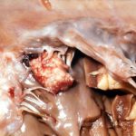
Image A. Multiple small lesions at both ends of the cordae tendinae in this patient who died of disseminated aspergillosis. He was a previously well 70 year old man who developed pneumonia on holiday, required artificial ventilation and died with a rapidly progressive cavitating pneumonia. Autopsy showed disseminated aspergillosis.
Image B. Another lesion in pt DB, that histologically showed a mass of hyphae and fibrin.
Image C. Large destructive lesion on the mitral valve in patient DB.
 ,
,  ,
, 
-
The patient was a 610 g twin male born by spontaneous normal vaginal delivery at 23 weeks and 4 days gestation. He was started on benzyl penicillin and gentamicin for sepsis. On day 3, he developed metabolic acidosis, hyponatremia, anemia, thrombocytopenia and jaundice and his antibiotics were changed to vancomycin, cefotaxime and fluconazole.
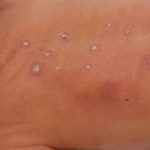
On day 10, multiple circular skin papules with white eschars were noted on his back (Figure A). A full septic screen was repeated including skin scraping and biopsy for urgent microscopy and culture. Microscopy of skin scrapes revealed fungal elements including hyphae and fruiting heads suggestive of Aspergillus spp (Figure B). Lipid amphotericin B was commenced and fluconazole was stopped. Skin scrapings on culture grew Aspergillus fumigatus. A diagnosis of primary cutaneous aspergillosis was made. The patient responded to oral posaconazole 6mg/kg/8 hourly. All lesions disappeared after 44 days and he continued with posaconazole until day 60.
Published case at Langan et al Pediatr Dermatol 2010 Jul-Aug 27 (4) 403-4
 ,
, 