Date: 26 November 2013
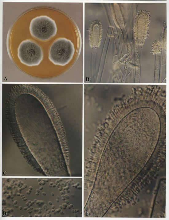
A Colonies on MEA after one week;B conidial heads and tip of conidiophore, x 230; C conidial head, x920; E conidial head, x920. Copyright B.Flannigan, R Samson & JD Miller (From Microorganisms in home and indoor work environments, Published by Taylor and Francis)
Copyright: n/a
Notes:
Aspergillus clavatus – A Colonies on MEA after one week;B conidial heads and tip of conidiophore, x 230; C conidial head, x920; E conidial head, x920. Copyright B.Flannigan, R Samson & JD Miller (From Microorganisms in home and indoor work environments, Published by Taylor and Francis)
Images library
-
Title
Legend
-
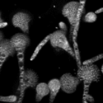
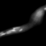
Mitochondria organisation: GFP fluorescence micrographs showing mitochondrial organisation in an A.nidulans strain with GFP mitochondria, grown at 25°C in minimal media.
-
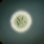
Colony morphology of A.nidulans SRF200 after two days at 37°C
-
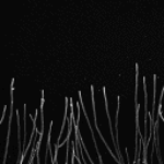
Aspergillus nidulans. Cell nuclei-Ds red. DsRed fluorescence micrographs showing nuclear distribution in an A.nidulans germling with dsRed stained nuclei

-
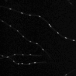
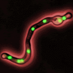
Cell Biology – Aspergillus nidulans. Cell nuclei-GFP. Nuclear distribution: GFP fluorescence mirographs showing fungal cell morphology and nuclear distribution in A.nidulans. GFP stained nuclei,grown at 25°C in minimal media O/N
-
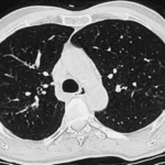
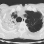
High resolution CT scan of chest.CT scan demonstrating remarkable bronchial wall thickening of the right main bronchus and main branches, in context of longstanding ABPA


