Date: 26 November 2013
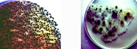
Left= an agar air plate exposed for 2 minutes after the barley had been turned. showing numerous colonies of the fungus following incubation at 26C on 2% malt agar.Right= A sputum sample taken from a maltworker after exposure showing many fungal colonies when cultured on agar. His commensal yeast flora is seen towards the right base as cream/white colonies.
Copyright: n/a
Notes: n/a
Images library
-
Title
Legend
-
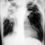
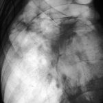
Right apical aspergilloma, patient WC. Plain chest radiograph of patient with right apical aspergilloma in an old, large tuberculous cavity. Severe haemoptysis and respiratory insufficiency together constituted the indications for embolisation which was done in one session over a 3 hour period (see images 1-6).
-
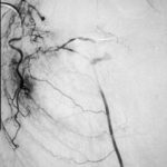
Embolisation 7 – patient WC. Angiogram of the lateral thoracic artery on subtraction film showing grossly abnormal vasculature inferiorly shunting along several anterior intercostal arteries to the internal mammary artery. In addition a pseudoaneurysm is shown.
-
Embolisation 6 – patient WC. Catheter tip in the lateral thoracic artery on screening film.
-
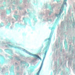
Grocott (silver) stain showing branching septate hyphae fairly typical of Aspergillus in mucus. The apparent right angle branching is unusual.
-
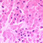
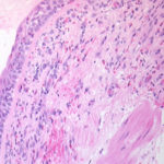
Bronchial mucosa under H & E stain showing numerous eosinophils deep to the mucosa, and mucus in the lumen of the bronchiole.
-
Grocott (silver) stain showing branching septate hyphae fairly typical of Aspergillus in mucus. The apparent right angle branching is unusual.

-
Severe kyphoscoliosis caused by greater than 40 years of prednisolone for ABPA and asthma.


