Date: 26 November 2013
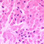
Aspergillosis in penguins. Lesions found in captive Magellanic penguins (Spheniscus magellanicus) with aspergillosis as determined by histology. Adherence areas of air sac to the celomic wall and white-yellowish nodule in the liver.
Copyright:
Kindly provided by Melissa Orzechowski Xavier [melissaxavier@bol.com.br], UFRGS – Brazil and with thanks to CRAM, Rio Grande, RS, Brazil.
Notes: n/a
Images library
-
Title
Legend
-
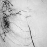
Embolisation 7 – patient WC. Angiogram of the lateral thoracic artery on subtraction film showing grossly abnormal vasculature inferiorly shunting along several anterior intercostal arteries to the internal mammary artery. In addition a pseudoaneurysm is shown.
-
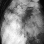
Embolisation 6 – patient WC. Catheter tip in the lateral thoracic artery on screening film.
-
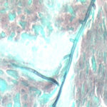
Grocott (silver) stain showing branching septate hyphae fairly typical of Aspergillus in mucus. The apparent right angle branching is unusual.
-
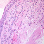
Bronchial mucosa under H & E stain showing numerous eosinophils deep to the mucosa, and mucus in the lumen of the bronchiole.
-
Grocott (silver) stain showing branching septate hyphae fairly typical of Aspergillus in mucus. The apparent right angle branching is unusual.

-
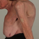
Severe kyphoscoliosis caused by greater than 40 years of prednisolone for ABPA and asthma.

-
These pictures show remarkable curvature of the spine as a result of collapse of the vertebral bodies of the thoracic vertebrae. This is a gross example of steroid-induced osteoporosis. The dose was not large in the last 10 years, typically 5-10mg daily, but multiple high dose courses and slow tapering lead to this outcome.
Her corticosteroid warning card is also demonstrated, as additional steroids are required for any significant illness or surgery, as her adrenal glands had completely atrophied.
Kindly supplied by Prof David Denning, South Manchester University Hospitals NHS Trust, Manchester UK
(© Fungal Research Trust)
 ,
,  ,
,  ,
,  ,
, 


