Date: 26 November 2013
History : A 20 yr old female with cystic fibrosis complicated by CF-related Diabetes mellitus (diagnosed at age 12 years) & ABPA (diagnosed at
15 years ). She was fairly stable until last 9 months, when she started to experience frequent pulmonary exacerbations, which have prompted intensive therapies.
Her serum IgE at the time of diagnosis was 5060 IU/L, skin prick test for aspergillus was positive, and serum was positive for precipitating antibodies to Aspergillus. She was treated with oral prednisone (1 mg/kg/day) for first two weeks followed by prednisone at 0.5 mg/kg every other day for at least 6 months with some clinical and serologic improvement. Over the following 5 years, she presented with a pattern of repeated episodic exacerbations with wheezing and crackles, increases in IgE and need to increase prednisone dosage. In an attempt to control her frequent ABPA relapses, itraconazole was added at 200mg twice a day, with some clinical & serologic improvement.
2-3 weeks prior to the scans and X rays, she developed severe protracted coughing spells associated with minor hemoptysis, low grade intermittent fever, and weight loss. Her FEV1 declined in a 3 months period from 56% to 33%.
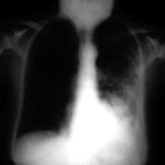
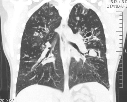
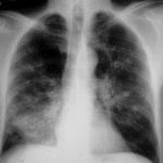
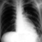
A chest-x ray (Fig 3) did not reveal any new changes when compared to the one obtained almost a year before. A CT scan of the chest Figs 1 and 2- lateral view), however shows an ovoid soft tissue density within an ectatic bronchi in the anterior basal segment of the LLL, felt to be an aspergilloma.
She was started on oral voriconazole (3 months after the above scans and X-rays) which is better absorbed in CF patients than itraconazole, at 200mg twice a day. Her prednisone was gradually reduced to 5 mgs/day and her FEV1 increased to 46% of predicted (03/31/10), her IgE level was 1167 kU/L (previously in January 2009, her IgE level was 3053 kU/L)and her weight has plateaued.
Since she was not fit enough for surgery at that time, removing the aspergilloma by flexible bronchoscopy was unsuccessful. The aspergilloma can be seen at bronchoscopy in Fig 4.
Go to follow up 12 months later
Copyright:
Kindly donated by Dr N Turcios, Director of pediatric pulmonology/cystic fibrosis in Somerville, NJ.
Notes: n/a
Images library
-
Title
Legend
-
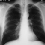
25/04/90 After itraconazole treatment. Major improvement, defined as a complete response, after 10 weeks therapy with itraconazole.
-
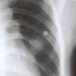
Image A. Chest x-ray shows a single nodule in the left mid lung field.
Image B. This emphasises how chest x-rays in this context underestimate the extent of disease. The most anterior nodule has ground glass surrrounding the nodule, a halo sign. This diagnostic feature is missed on plain chest X-rays.
 ,
, 
-
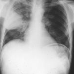
Chest X ray after 4 days, prior to treatment, showing massive increase in volume of lesion (Fig 2)
-
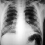
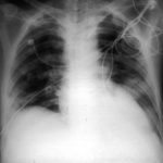
Image A. This patient, aged 25 years developed a non productive cough and dyspnoea in the context of late-stage AIDS, CMV disease with ganciclovir-induced neutropenia and receiving corticosteroids. His chest radiograph shows fine bilateral reticular lower-lobe shadowing. He then developed gastro-intestinal bleeding with a gastric ulcer which showed hyphae on biopsy. He then developed blindness of one eye and the globe of his eye perforated. Hyphae were seen and Aspergillus cultured from the vitreous aspirate.
Image B. This radiograph, taken 25 days after the first and 3 days before death, shows of fine bilateral lower-lobe reticular shadows progressing to nodules in all lung zones.
This patient was reported as patient 3 in Denning DW, Follansbee S, Scolaro M, Norris S, Edelstein D, Stevens DA. Pulmonary aspergillosis in the acquired immunodeficiency syndrome. N Engl J Med 1991; 324: 654-662.
 ,
, 
-
Further details
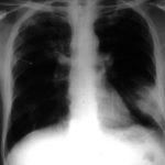
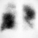
Image A. Bronchoscopy revealed Aspergillus on culture.
Image B. The ability of Aspergillus to cause pulmonary infarction, probably through direct angioinvasion in this case, is characteristic.
 ,
, 
-
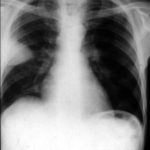
(Fig 1) Chest radiograph with ‘classical’ appearance of a pulmonary infarction – a wedge-shaped lesion peripherally set against the pleura.
-
Large soft left upper-lobe shadow of focal invasive pulmonary aspergillosis in leukaemia, that was missed on earlier radiographs but apparent retrospectively. Variable density of the lesion suggests cavitation, which would be clearly visible on a CT scan of the thorax.
-
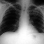
Severe unilateral invasive aspergillosis of the left lung, with complete consolidation of the left lower-lobe and reticular shadowing extending up into the left upper lobe. The right lung appears normal.
