Date: 26 November 2013
Further image details
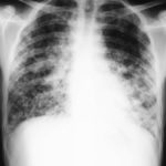
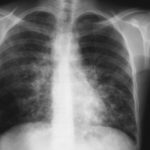
Image A. Long standing sarcoidosis, on corticosteroids with fibrosis and cavitary disease, and a possible fungal ball in the cavity on the left (1996).
Image B. Long standing sarcoidosis, on corticosteroids with 2 cavities containing aspergillomas, one on the left and one on the right (1996).
Image C. Sarcoidosis with progressive cavity formation and aspergillomas. Probable CIPA given appearances (2000).
Image D. Sarcoidosis with progressive cavity formation and aspergillomas. Probable CIPA given appearances (2000).
Image E. Sarcoidosis with progressive cavity formation and aspergillomas. Probable CIPA given appearances (2000).
Copyright: n/a
Notes: n/a
Images library
-
Title
Legend
-
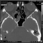
Allergic Aspergillus Sinusitis -Patient AM. D – Marked involvement of ehmoidal air cells on the right , together with the inferior aspect of the sphenoid sinus. The left side is almost clear of disease.
-
Further details
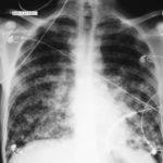
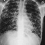
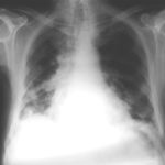
Image 1. The chest x-ray shows extensive bilateral nodular disease, most consistent with a fungal infection, or possibly tuberculosis. He was treated with a bucket face mask with 80% oxygen and voriconazole.
 ,
,  ,
,  ,
,  ,
,  ,
, 
-
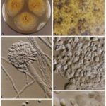
A Colonies on MEA +20 % sucrose after 2 weeks; B ascomata, x 40; C conidiophore of Aspergillus glaucus x 920;D conidiophore of Aspergillus glaucus x920 E. portion of ascoma with asci x 920. F ascospores x2330.
-
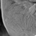
Scanning electron micrographs of A. fumigatus conidia of transformants rodB-02 (b). Size bar, 100 nm.

-
Scanning electron micrographs of A. fumigatus conidia of the wild-type G10 strain (a). Size bar, 100 nm.
-
Scanning electron micrographs of A. fumigatus conidia of rodA rodB-26 (d).Size bar, 100 nm.

 ,
, 
