Date: 26 November 2013
Further image details
Image A. Long standing sarcoidosis, on corticosteroids with fibrosis and cavitary disease, and a possible fungal ball in the cavity on the left (1996).
Image B. Long standing sarcoidosis, on corticosteroids with 2 cavities containing aspergillomas, one on the left and one on the right (1996).
Image C. Sarcoidosis with progressive cavity formation and aspergillomas. Probable CIPA given appearances (2000).
Image D. Sarcoidosis with progressive cavity formation and aspergillomas. Probable CIPA given appearances (2000).
Image E. Sarcoidosis with progressive cavity formation and aspergillomas. Probable CIPA given appearances (2000).
Copyright: n/a
Notes: n/a
Images library
-
Title
Legend
-
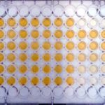
Test for aflatoxin B1. Standard curve wells in triplicate – columns 9, 10 & 11 – laid out horizontally. Triplicates of 14 samples laid out vertically in rows 2 to 8
-
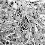
Light microscopical appearance of hepatic tissue in outbreak of aflatoxicosis showing centrilobular degeneration of hepatocytes with vacuolated cytoplasm and apoptoses (top right abnormal)(Haematoxylin and eosin).
-
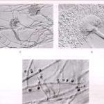
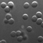
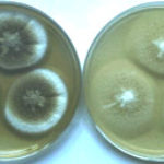
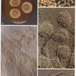
Aspergillus terreus Thom. A Colonies on MEA after one week; B detail of colony showing columnar conidial heads x 44 ; C conidial heads and tip x 920; D conidia x2330
-
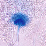
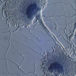
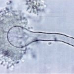
Aspergillus terreus Thom. Conidial head of Aspergillus terreus. Conidial heads are compact, columnar and biseriate. Conidiophores are hyaline to slightly yellow and smooth walled.
