Date: 26 November 2013
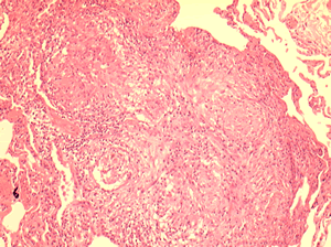
Allergic Bronchocentric Granulomatosis. Low power. Sections show muscle, lung with acute inflammation and evidence of organisation with early fibrosis. The bronchial wall can be seen with chronic inflammation and many eosinophils.There is a thickened basement membrane. No definite granulomata are seen.
Copyright: n/a
Notes:
A 53 year old male with fevers, shortness of breath and a progressive left lower lobe infiltrate, he had a previous history of aspergillosis. A percutaneous lung biopsy was done. All sections were stained with haemotoxylin and eosin. No fungal hyphae were seen with silver staining (not shown).
Images library
-
Title
Legend
-
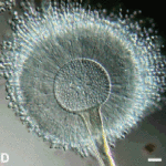
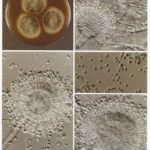
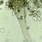
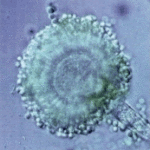
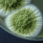
Aspergillus flavus Link. A colonies after 1 weekB,C conidial heads x 920D Conidia x920 E Conidial head x920
-
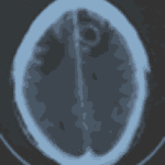
Patient diagnosed with Stage C Chronic Lymphocytic Leukaemia, treated in the MRC CLL4 study, with prednisolone for 4 weeks followed by oral chlorambucil for 7 days. Patient developed severe pneumonia due to pseudomonas and staphylococcus. Following treatment with broad spectrum antibiotics and 1 week of Abelcet, patient was readmitted with headache, disorientation and fever. CT brain scans showed 3 ring enhancing lesions, aspirated material showed neutrophils but grew aspergillus. Patient now improved on Abelcet (4 mg/kg) and oral itrconazole suspension 200mg b.d.
Thanks to Richard Chasty, Consultant Haematologist, North Staffordsire Hospital, UK.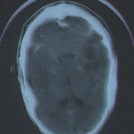 ,
, 

![Asp[1]niger Asp[1]niger](https://www.aspergillus.org.uk/wp-content/uploads/2013/11/Asp1niger-150x150.gif)


