Date: 26 November 2013
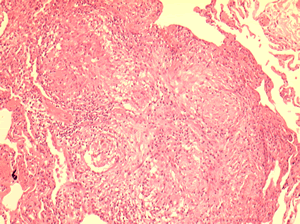
Allergic Bronchocentric Granulomatosis. Low power. Sections show muscle, lung with acute inflammation and evidence of organisation with early fibrosis. The bronchial wall can be seen with chronic inflammation and many eosinophils.There is a thickened basement membrane. No definite granulomata are seen.
Copyright: n/a
Notes:
A 53 year old male with fevers, shortness of breath and a progressive left lower lobe infiltrate, he had a previous history of aspergillosis. A percutaneous lung biopsy was done. All sections were stained with haemotoxylin and eosin. No fungal hyphae were seen with silver staining (not shown).
Images library
-
Title
Legend
-
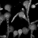
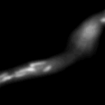
Mitochondria organisation: GFP fluorescence micrographs showing mitochondrial organisation in an A.nidulans strain with GFP mitochondria, grown at 25°C in minimal media.
-
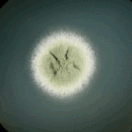
Colony morphology of A.nidulans SRF200 after two days at 37°C
-
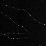
Aspergillus nidulans. Cell nuclei-Ds red. DsRed fluorescence micrographs showing nuclear distribution in an A.nidulans germling with dsRed stained nuclei

-
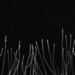
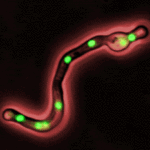
Cell Biology – Aspergillus nidulans. Cell nuclei-GFP. Nuclear distribution: GFP fluorescence mirographs showing fungal cell morphology and nuclear distribution in A.nidulans. GFP stained nuclei,grown at 25°C in minimal media O/N
-
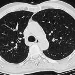
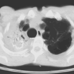
High resolution CT scan of chest.CT scan demonstrating remarkable bronchial wall thickening of the right main bronchus and main branches, in context of longstanding ABPA


