Date: 26 November 2013
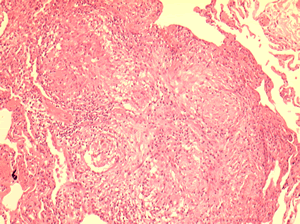
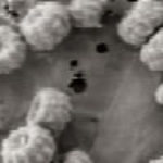
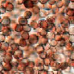
Allergic Bronchocentric Granulomatosis. Low power. Sections show muscle, lung with acute inflammation and evidence of organisation with early fibrosis. The bronchial wall can be seen with chronic inflammation and many eosinophils.There is a thickened basement membrane. No definite granulomata are seen.
Copyright: n/a
Notes:
A 53 year old male with fevers, shortness of breath and a progressive left lower lobe infiltrate, he had a previous history of aspergillosis. A percutaneous lung biopsy was done. All sections were stained with haemotoxylin and eosin. No fungal hyphae were seen with silver staining (not shown).
Images library
-
Title
Legend
-
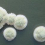
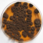
Pigmentation of Aspergillus versicolor colonies ranged from pale green to greenish-beige, pink-green, dark green and brown. Reverse is usually reddish. The growth rate is usually slow. Cultured on Sabouraud dextrose agar with chloramphenicol.
-
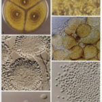
A Colonies on MEA after one week; B, C conidial heads with tip of conidiophire, x920; D conidial head, x 2330; E conidial heads x920
![aspvers[2] aspvers2](https://www.aspergillus.org.uk/wp-content/uploads/2017/10/aspvers2-150x150.jpg)
-
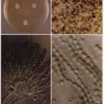
A Colonies on MEA + 20% sucrose after one week; B detail of colony showing columnar conidial heads x 44 ; C conidial heads x 920; D conidia x2330
-
Cultures are grown on malt extract agar for 5-7 days at 30°C.
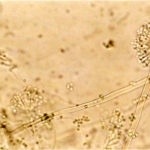
Light microscopy-1000x stained with lacto-phenol and cotton blue.
-
A Colonies on MEA +20% sucrose after one week; B ascomata x 40; C conidiophores x 920; D ascospores x2330; E ascoma x 230; F portion of ascoma with asci and ascospores, x 920.
