Date: 26 November 2013
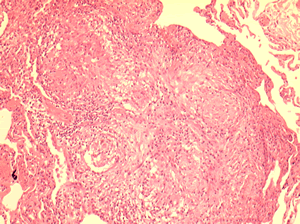
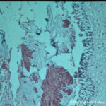
Allergic Bronchocentric Granulomatosis. Low power. Sections show muscle, lung with acute inflammation and evidence of organisation with early fibrosis. The bronchial wall can be seen with chronic inflammation and many eosinophils.There is a thickened basement membrane. No definite granulomata are seen.
Copyright: n/a
Notes:
A 53 year old male with fevers, shortness of breath and a progressive left lower lobe infiltrate, he had a previous history of aspergillosis. A percutaneous lung biopsy was done. All sections were stained with haemotoxylin and eosin. No fungal hyphae were seen with silver staining (not shown).
Images library
-
Title
Legend
-
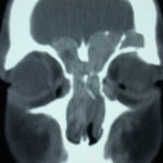
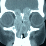
4 Total obstruction of the sinuses due to inflamed mucosa. (Patient 04)
-
1 Axial computed tomography (CT) scans of the frontal sinus.
A: due to the long lasting pressure of mucus, the bone of the anterior wall of frontal sinus is thinned out and elevated anteriorly, forming a bulge. B: same situation as depicted in fig A: the posterior bony wall of frontal sinus is thinned out and extremely elevated posteriorly towards the frontal lobe of the brain. As depicted on the scan, a thin bony layer covering the dura could be recognized intraoperatively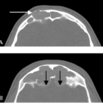
-
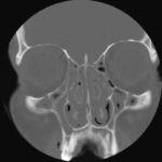
2 Same patient as 1 and 3, frontal CT
-
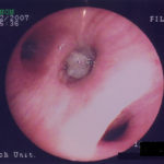
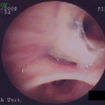
D. 6 months later, tenacious yellow secretions in L basal bronchial division
-
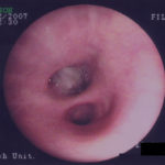
C. After suction the material was seen to extend distally – obstructing the right basal stem bronchus
-
B. After suction the material was seen to extend distally – obstructing the right basal stem bronchus
