Date: 26 November 2013
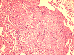
Allergic Bronchocentric Granulomatosis. Low power. Sections show muscle, lung with acute inflammation and evidence of organisation with early fibrosis. The bronchial wall can be seen with chronic inflammation and many eosinophils.There is a thickened basement membrane. No definite granulomata are seen.
Copyright: n/a
Notes:
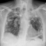
A 53 year old male with fevers, shortness of breath and a progressive left lower lobe infiltrate, he had a previous history of aspergillosis. A percutaneous lung biopsy was done. All sections were stained with haemotoxylin and eosin. No fungal hyphae were seen with silver staining (not shown).
Images library
-
Title
Legend
-
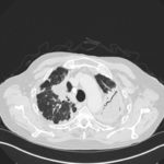
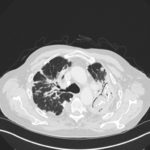
Mr RM is 80 and an ex-coal miner.He developed pneumoconiosis from exposure to coal dust. He also developed rheumatoid arthritis and the combination of this disease and pneumoconiosis is called Caplan’s syndrome.
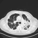
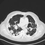
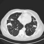
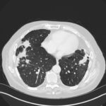
His chest Xray in early 2015 shows extensive bilateral pulmonary shadowing with solid looking nodules superimposed on abnormal lung fields, contraction of his left lung with an elevated diaphragm and a large left upper lobe aspergilloma, displaying a classic air crescent. His CT scan from mid 2014 demonstrates a large aspergilloma in a cavity on the left, with marked pleural thickening around it, which is partially ‘calcified’ towards its base. Inferiorly on other images,remarkable pleural thickening and fibrotic irregular and spiculated nodules are seen, most partially calcified.
 ,
,  ,
,  ,
,  ,
,  ,
,  ,
, 