Date: 3 February 2014
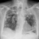
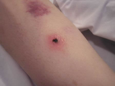
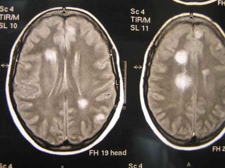
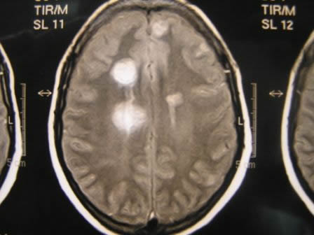
This patient was 28 yr old with adult lymphocytic leukaemia. She received induction chemotherapy and this infection developed 2 days after recovering from neutropenia.
Copyright:
These images were kindly donated by Maria Cecilia Dignani, MD, Head Infectious Diseases, Fundaleu, Buenos Aires, Argentina. September 2007
(© Fungal Research Trust)
Notes:
A,B,&C the patient had multiple erythematous lesions on the skin, some of the lesions were papular and showed necrotic tissue.
D,E,F & G MRI brain scans showed multiple bilateral nodular lesions in the frontal, parietal and occipital areas. Lesions were 0.7 – 2cm in diameter involving both peripheral and central areas of the brain. G– illustrates a small well defined lesion in the posterior fossa.
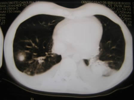
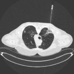
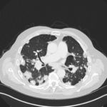
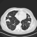
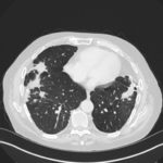
H,I & J CT scans of lungs showing nodular lesions in both lungs with infiltrates. I – exhibits the classic halo sign – suggestive of aspergillus infection. J illustrates that some associated pleural effusion was present.
The patient was exposed to high levels of mould during treatment for ALL as a result of a leaking pipe and extensive mould damage to wallcoverings in her home.
Images library
-
Title
Legend
-
Mr RM is 80 and an ex-coal miner.He developed pneumoconiosis from exposure to coal dust. He also developed rheumatoid arthritis and the combination of this disease and pneumoconiosis is called Caplan’s syndrome.
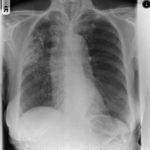
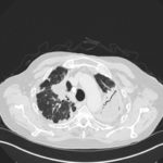
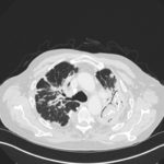
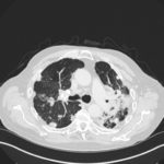
His chest Xray in early 2015 shows extensive bilateral pulmonary shadowing with solid looking nodules superimposed on abnormal lung fields, contraction of his left lung with an elevated diaphragm and a large left upper lobe aspergilloma, displaying a classic air crescent. His CT scan from mid 2014 demonstrates a large aspergilloma in a cavity on the left, with marked pleural thickening around it, which is partially ‘calcified’ towards its base. Inferiorly on other images,remarkable pleural thickening and fibrotic irregular and spiculated nodules are seen, most partially calcified.
 ,
,  ,
,  ,
,  ,
,  ,
,  ,
, 