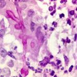
Date: 26 November 2013
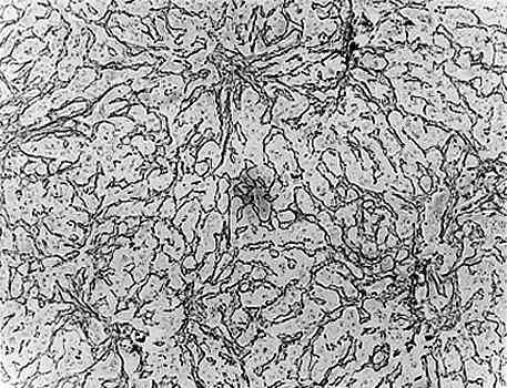
Light microscopical appearance of hepatic tissue in outbreak of aflatoxicosis showing centrilobular collapse with reticular condensation (Reticulin).
Copyright:
© Fungal Research Trust
Notes:
Ref.: Ngindu, A., et al., Outbreak of acute hepatitis caused by aflatoxin poisoning in Kenya. Lancet 1982; 1: 1346-48.
Images library
-
Title
Legend
-
BAL specimen showing hyaline, septate hyphae consistent with Aspergillus, stained with Blankophor
-
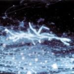
Mucous plug examined by light microscopy with KOH, showing a network of hyaline branching hyphae typical of Aspergillus, from a patient with ABPA.
-
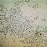
Corneal scraping stained with lactophenol cotton blue showing beaded septate hyphae not typical of either Fusarium spp or Aspergillus spp, being more consistent with a dematiceous (ie brown coloured) fungus
-
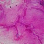
Corneal scrape with lactophenol cotton blue shows separate hyphae with Fusarium spp or Aspergillus spp.
-
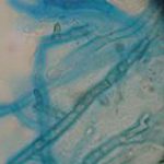
A filamentous fungus in the CSF of a patient with meningitis that grew Candida albicans in culture subsequently.
-
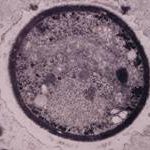
Transmission electron micrograph of a C. neoformans cell seen in CSF in an AIDS patients with remarkably little capsule present. These cells may be mistaken for lymphocytes.
-
India ink preparation of CSF showing multiple yeasts with large capsules, and narrow buds to smaller daughter cells, typical of C. neoformans
