Date: 26 November 2013
Copyright: n/a
Notes: n/a
Images library
-
Title
Legend
-
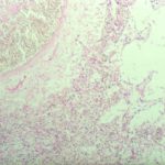
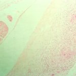
Pulmonary aspergillosis (cow 2). Taken from the edge of a lesion, as described in the previous image for cow 2.
-
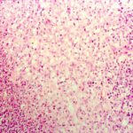
Pulmonary aspergillosis (cow 2). Section of lung from a 2 year old cow with weight loss and anorexia since calving. On necropsy examination, multiple firm masses were identified throughout the lungs. These were cavitating in nature, with a necrotic centre and peripheral fibrosis. Both this section and the following one are taken from the edge of such a lesion and demonstrate the pyogranulomatous inflammatory response.
-
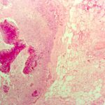
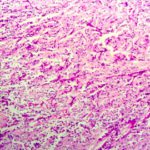
Guttural pouch aspergillus (PAS) (horse TW). Serial section of previous slide for horse TW stained by PAS reveals the presence of fungal hyphae within the inflammatory tissue. Aspergillus spp. is the likely cause of equine guttural pouch mycosis.
-
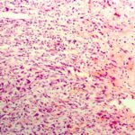
Guttural pouch aspergillosis (horse TW). Section of the wall of the guttural pouch from an 8 year old, neutered male horse with recurrent right-sided epistaxis of 10 days duration. Endoscopic examination confirmed the origin of the blood to be the guttural pouch and the right internal carotid artery was ligated surgically. Necropsy examination revealed an extensive ruptured aneurysm of this vessel and guttural pouch mycosis. The section reveals necrosis and chronic inflammatory infiltration of guttural pouc
-
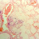
Pulmonary aspergillosis. Section of lung from a 6 year old, Holstein cow with mycotic pneumonia attributed to Aspergillus spp.
-
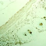
Meningitis (dog R). Grocott-stained serial section of the previous slide for dog R reveals the presence of fungal material at the centre of each microgranuloma. These lesions were not cultured, but Aspergillus spp. was identified by immunohistochemical examination using a panel of specific antisera. There was no evidence of systemic involvement in this dog.
-
Aspergillus meningitis (dog R). Section of spinal meninges from a 4 year old, neutered female German shepherd dog with multiple discospondylitis involving T1-2, T6-7, T10-11 and L5-6. There are a series of coalescing microgranulomas associated with meninges at each site.
-
Pulmonary aspergillosis (cat T). Section of lung from a young adult, female domestic short haired cat. This was one of seven unvaccinated farm cats that died over a 12 month period. At necropsy examination there was diffuse pulmonary consolidation with scattered abscessation. Aspergillus spp. was cultured from these lesions. The cat also had histopathological evidence of lymph node atrophy, a change that may be attributed to feline immunodeficiency virus (FIV) infection.

-
Pulmonary aspergillosis (PAS) (parrot C). Section of lung from parrot C stained by periodic acid schiff (PAS) demonstrating the hyphal material. Aspergillus spp and Bacillus cereus were cultured from the lesions.
