Date: 26 November 2013
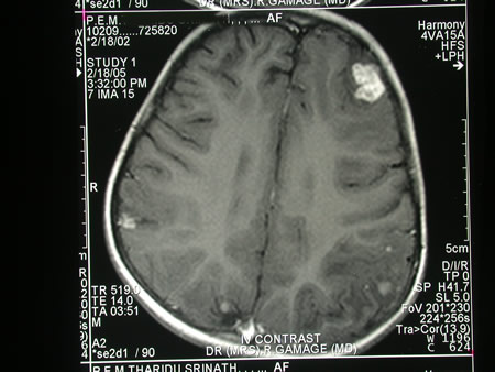
Image c. 3 yr old boy with CNS aspergillosis pt TS. MRI scan pre-amphotericin B
Copyright: n/a
Notes:
A 3 year old boy, quite active and healthy clinically, who has CNS aspergillosis. He was first seen about 4 months ago for a red eye, which turned out to be panophthalmitis; culture yielded Aspergillus spp. He received 2 weeks of iv amphotericin and was sent home by the ophthalmologists. No h/o eye trauma. He returned 2 weeks ago with focal fits, and the MR showed several lesions bilaterally (including ring enhancing lesions) and normal sinuses, and a brain bx showed fungal hyphae (no culture this time). His immune status (normal WCC and neutrophil function so far) was investigated.
He was given conventional amphotericin for 8 weeks, and switched to oral itraconazole. We had to limit the ampho to 0.7 mg/kg owing to toxicity (mainly hypokalaemia).
The MRI scan was repeated at about 6 weeks, and generally showed good improvement (scans e-h). The enhancement/flare were gone but remained in a few lesions, the lesions themselves were all either gone or much smaller. Further investigations revealed the child was immunocompetent.
Patient was switched from amphotericin to oral itraconazole at week 8 essentially on a clinical assessment. Awaiting follow-up.
Images library
-
Title
Legend
-
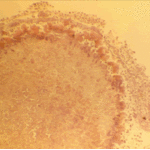
The periphery of the fungus ball is deeply eosinophilic because of the deposition of Splendore-Hoeppli material.
-
Single fungal ball, moving. Radiographic appearance of a fungus ball, showing movement as the patient’s position changes.
-
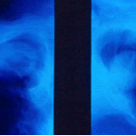
Oxalate crystals in the cavity wall surrounding an Aspergillus niger fungus ball (H&E, dark field, x 25).
-
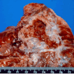
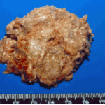
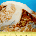
Aspergilloma patient. Gross pathology appearance of a fungus ball.
-
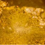
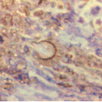
Conidiophores of Aspergillus fumigatus in the mass of the fungal ball surrounded by mycelia (H&E, x 400).
-
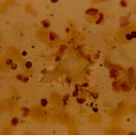
Aspergillus niger fungal ball. Calcium oxalate crystals in Aspergillus niger fungal ball. Also shown are darkly pigmented, rough-walled conidia associated with Aspergillus niger infection.
-
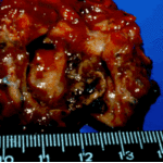
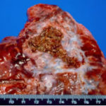
Aspergillus niger fungus ball within an old tuberculous cavern. This patient had diabetes, a disease commonly associated with A. niger infection.

 ,
, 
 ,
, 