Date: 26 November 2013
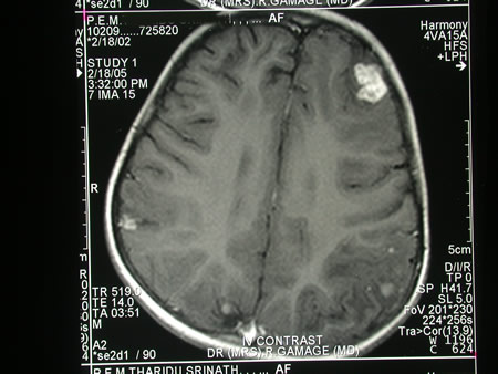
Image c. 3 yr old boy with CNS aspergillosis pt TS. MRI scan pre-amphotericin B
Copyright: n/a
Notes:
A 3 year old boy, quite active and healthy clinically, who has CNS aspergillosis. He was first seen about 4 months ago for a red eye, which turned out to be panophthalmitis; culture yielded Aspergillus spp. He received 2 weeks of iv amphotericin and was sent home by the ophthalmologists. No h/o eye trauma. He returned 2 weeks ago with focal fits, and the MR showed several lesions bilaterally (including ring enhancing lesions) and normal sinuses, and a brain bx showed fungal hyphae (no culture this time). His immune status (normal WCC and neutrophil function so far) was investigated.
He was given conventional amphotericin for 8 weeks, and switched to oral itraconazole. We had to limit the ampho to 0.7 mg/kg owing to toxicity (mainly hypokalaemia).
The MRI scan was repeated at about 6 weeks, and generally showed good improvement (scans e-h). The enhancement/flare were gone but remained in a few lesions, the lesions themselves were all either gone or much smaller. Further investigations revealed the child was immunocompetent.
Patient was switched from amphotericin to oral itraconazole at week 8 essentially on a clinical assessment. Awaiting follow-up.
Images library
-
Title
Legend
-
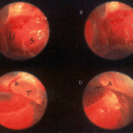
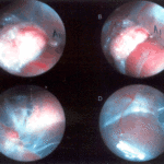
Falcons: The following images were obtained by endoscopy of falcons with aspergillosis.A,B Thoracic airsac (T) with prominent blood vessels and a dead serratospiculum worm (W). The presence of these lung worms makes the airsac look milky. D Normal ovary with developing follicles.

-
Falcons: The following images were obtained by endoscopy of falcons with aspergillosis.B,D Aspergillus lesions (A) over a swollen liver

-
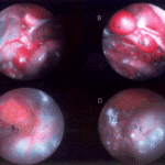
Falcons: The following images were obtained by endoscopy of falcons with aspergillosis.B Cranial, middle, caudal lobes (K1,K2,K3) of the left kidney, all the lobes show slight nephromegaly.C Yellow aspergillus colony (A1), lying adjacent to the lung.D White aspergillus colonies (A2,A3,A4).
-
Falcons: The following images were obtained by endoscopy of falcons with aspergillosis.C Cranial pole of left kidney (K) -mildly inflamed.D Ovary ( F) with developing follicles.
-
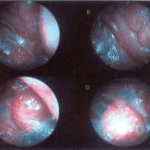
The following images were obtained by endoscopy of falcons with aspergillosis.A and B Lung Worm (S) over liver (Li) (serratospiculum seurati)C and D Aspergilloma (A) and prominent blood vessels on the caudal thoracic air sac (T).
